Teaching NeuroImages: Herpes zoster ophthalmicus–related oculomotor palsy accompanied by Hutchinson sign
Gayatri S. Reilly and Robert K. Shin
Neurology. April 13, 2010; 74 (15) RESIDENT AND FELLOW SECTION
ARTICLE
A 51-year-old woman presented with acute diplopia. Findings include right ptosis, a dilated, unreactive pupil, and impaired adduction and vertical ductions (figure 1). A skin lesion was noted on the right tip of the nose, residual from a vesicular rash over the right forehead 3 weeks earlier (figure 1). MRI demonstrated enhancement of the cisternal third nerve, obviating the need for angiography (figure 2). The oculomotor palsy resolved within 3 months.
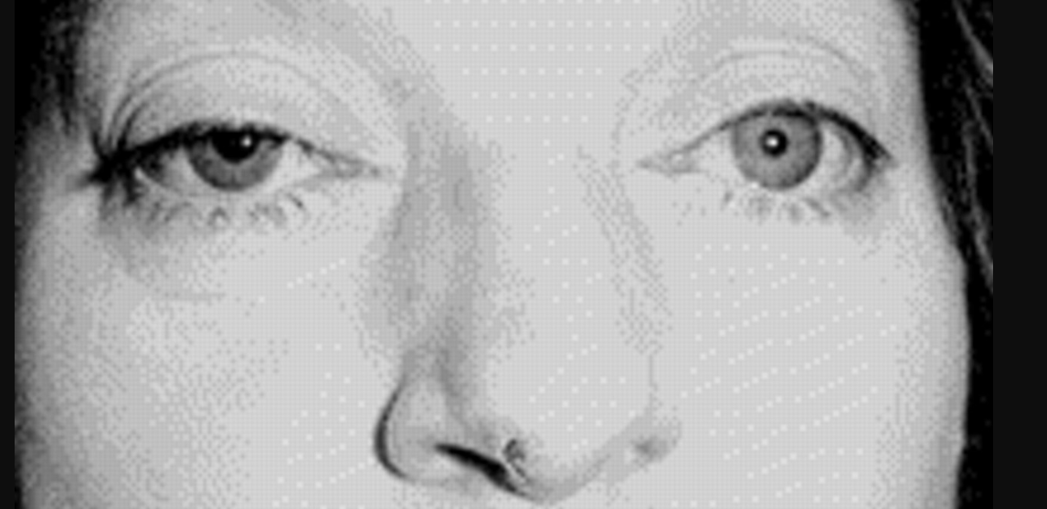
Figure 1 Partial ptosis, mydriasis, and exotropia consistent with a right oculomotor palsy
The skin lesion on the tip of the nose (Hutchinson sign) signifies involvement of the nasociliary branch of V1, which also innervates ocular structures.