Neuro-ophthalmology Illustrated Chapter 19 – Diagnosis of Headache and Facial Pain 2

Questions:
7. What differentiates classic from symptomatic trigeminal neuralgia?
8. Name several triggers for trigeminal neuralgia?
9. For Classic Trigeminal Neuralgia, what are the:
1. Age range
2. Gender ratio
3. Location of pain
4. Type of pain
5. Duration of pain
6. Temporal pain profile
7. Associated signs
8. Interictal exam
9. Treatment
10. For Symptomatic Trigeminal Neuralgia, what are the:
1. Age range
2. Gender ratio
3. Location of pain
4. Type of pain
5. Duration of pain
6. Temporal pain profile
7. Associated signs
8. Interictal exam
9. Treatment
11. For Cluster (Histamine) Headache, what are the: 
1. Age range
2. Gender ratio
3. Location of pain
4. Type of pain
5. Duration of pain
6. Temporal pain profile
7. Associated signs
8. Interictal exam
9. Treatment
12. For Episodic or Chronic Paroxysmal Hemicrania, what are the:
1. Age range
2. Gender ratio
3. Location of pain
4. Type of pain
5. Duration of pain
6. Temporal pain profile
7. Associated signs
8. Interictal exam
9. Treatment
13. For SUNCT Syndrome (Short-lasting, Unilateral, Neuralgiform headache attacks with Conjunctival injection and Tearing), what are the:
1. Age range
2. Gender ratio
3. Location of pain
4. Type of pain
5. Duration of pain
6. Temporal pain profile
7. Associated signs
8. Interictal exam
9. Treatment
14. For Hemicrania Continua, what are the:
1. Age range
2. Gender ratio
3. Location of pain
4. Type of pain
5. Duration of pain
6. Temporal pain profile
7. Associated signs
8. Interictal exam
9. Treatment

____________________________________________________

Questions with answers:
7. What differentiates classic from symptomatic trigeminal neuralgia?
The classic form is to be considered idiopathic. It includes the cases that are due to a normal artery in contact with the 5th nerve, such as the superior cerebellar artery or even a primitive trigeminal artery. The symptomatic form is not idiopathic and can have multiple origins: aneurysms, tumors, chronic meningeal inflammation, or other lesions may irritate trigeminal nerve roots along the pons. An abnormal vascular course of the superior cerebellar artery is often cited as the cause.

8. Name several triggers for trigeminal neuralgia?
1. Chewing, talking or smiling
2. Drinking cold or hot liquids
3. Touching, shaving, teeth brushing, nose-blowing 
4. Drafts of cold air 

9. For Classic Trigeminal Neuralgia, what are the:
1. Age range: 50-year-old
2. Gender ratio: 1/4
3. Location of pain: Unilateral V2 & V3 > V1
4. Type of pain: Stabbing
5. Duration of pain: <1 second
6. Temporal pain profile: Few to many/day for weeks to months. Pain-free between attacks.
7. Associated signs: No associated signs, look for triggers
8. Interictal exam: Normal
9. Treatment: Carbamazepine (Tegretol) frequent good response

10. For Symptomatic Trigeminal Neuralgia, what are the:
1. Age range: Any
2. Gender ratio: M=F
3. Location of pain: May be bilateral, V1, 2 or 3
4. Type of pain: Dull, persistent
5. Duration of pain: Chronic
6. Temporal pain profile: Fluctuations but no remission
7. Associated signs: 5th nerve hypoesthesia, dysesthesia, and motor deficit
and other cranial nerve palsies 
8. Interictal exam: 5th nerve hypoesthesia and motor deficit
9. Treatment: Variable response to therapy

11. For Cluster (Histamine) Headache, what are the: 
1. Age range: Young
2. Gender ratio: M/F 9/1
3. Location of pain: Unilateral periorbital
4. Type of pain: Boring, very severe
5. Duration of pain: 15-180 minutes
6. Temporal pain profile: 1-8/day, attack phase 4-16 weeks, Remission 6-24 months 
7. Associated signs: Horner syndrome, eye redness, tearing and rhinorrhea
8. Interictal exam: Normal but may have Horner syndrome
9. Treatment: Acute: oxygen, injectable triptans, and DHE, Chronic: lithium, steroids, channel inhibitors-Verapamil (Calan)

12. For Episodic or Chronic Paroxysmal Hemicrania, what are the:
1. Age range: any
2. Gender ratio: M/F 1/3
3. Location of pain: Unilateral periorbital
4. Type of pain: Very severe
5. Duration of pain: 2-30 minutes
6. Temporal pain profile: 5/day
7. Associated signs: Horner syndrome, eye redness, tearing and rhinorrhea
8. Interictal exam: Normal but may have Horner syndrome
9. Treatment: Indomethacin stops pain 

13. For SUNCT Syndrome (Short-lasting, Unilateral, Neuralgiform headache attacks with Conjunctival injection and Tearing), what are the:
1. Age range: Any
2. Gender ratio: M/F 8/1
3. Location of pain: Unilateral periorbital
4. Type of pain: Very severe
5. Duration of pain: 5-240 seconds
6. Temporal pain profile: Numerous in a day, sometimes dull pain between attacks, remissions with an irregular pattern
7. Associated signs: Eye redness and tearing
8. Interictal exam: Normal
9. Treatment: None, variable response (Corticosteroids and the antiepileptic drugs gabapentin, lamotrigine, and carbamazepine may help relieve some symptoms)

14. For Hemicrania Continua, what are the:
1. Age range: Any
2. Gender ratio: M>F
3. Location of pain: Unilateral hemicrania, always same side
4. Type of pain: Sharp pain
5. Duration of pain:  <1 minute
6. Temporal pain profile: Associated with a continuous headache of fluctuating intensity
7. Associated signs: Nausea, vomiting, photophobia and may have Horner syndrome
8. Interictal exam: Normal
9. Treatment: Indomethacin

____________________________________________________

The information below is from Neuro-ophthalmology Illustrated-2nd Edition. Biousse V and Newman NJ. 2012. Thieme

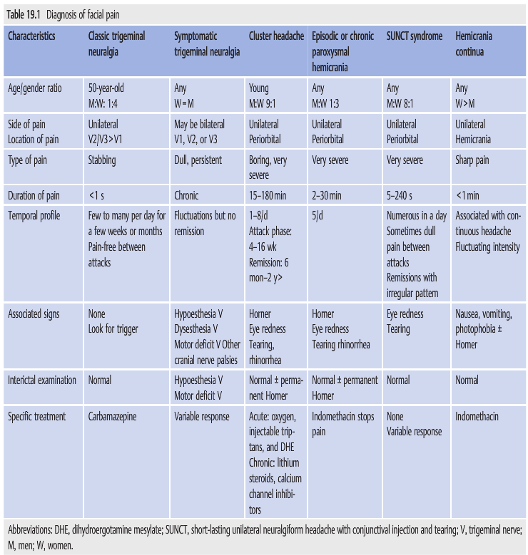
19.4 Differential Diagnosis of Facial Pain
▶Table 19.1 outlines the characteristics of various facial pain syndromes: classic trigeminal neuralgia, symptomatic trigeminal neuralgia, cluster headache, episodic or chronic paroxysmal hemicrania, SUNCT syndrome, and hemicrania continua. In most cases with new-onset facial pain or hemicrania, a workup is required to rule out underlying lesions (e.g., dissections, aneurysms, or cavernous sinus or skull base lesions).

Reference: 1. Neuro-ophthalmology Illustrated-2nd Edition. Biousse V and Newman NJ. 2012. Thieme

These questions are archived at https://neuro-ophthalmology.stanford.edu
Follow https://twitter.com/NeuroOphthQandA to be notified of new neuro-ophthalmology questions of the week.
Please send feedback, questions, and corrections to tcooper@stanford.edu.

Neuro-ophthalmology Illustrated Chapter 20 – Conditions Commonly Encountered in Neuro-ophthalmology 5

Questions:
38. What type of iris nodules are present in Neurofibromatosis Type 1, and  Neurofibromatosis Type 2?
39. What are the ocular or CNS manifestations of Neurofibromatosis Type 1?
40. What are the ocular or CNS manifestations of Neurofibromatosis Type 2?
41. What is the classic findings triad in tuberous sclerosis?
42. What is the most prominent ocular manifestation of Tuberous sclerosis?
43. What autosomal dominant condition is associated with multiple, bilateral retinal angiomas and intracranial cerebellar hemangioblastomas?
44. What condition should be suspected in a patient with findings of telangiectasias of the conjunctival vessels and oculomotor apraxia?
45. A patient has an upper eyelid hemangioma, intraocular hypertension, and homonymous hemianopia, what condition is likely to be present?
46. What are the ocular manifestations of Sturge-Weber syndrome?
47. What are the findings of the Wyburn-Mason Syndrome?
48. What are the findings of the Klippel-Trenaunay-Weber Syndrome?

Neuro-ophthalmology Illustrated Chapter 19 – Diagnosis of Headache and Facial Pain 3

Questions:
15. What should be measured in all patients with headache?
16. What must be ruled out in all patients with headache?
17. What must be measured in all patients with recurrent, unilateral pain localized around the eye?
18. What lab tests should be done in all patients over age 50 with any headache or facial pain?
19. What should be considered and done when a patient presents with a thunderclap headache? 

Neuro-ophthalmology Illustrated Chapter 19 – Diagnosis of Headache and Facial Pain 2

Questions:
7. What differentiates classic from symptomatic trigeminal neuralgia?

8. Name several triggers for trigeminal neuralgia?

9. For Classic Trigeminal Neuralgia, what are the:
1. Age range
2. Gender ratio
3. Location of pain
4. Type of pain
5. Duration of pain
6. Temporal pain profile
7. Associated signs
8. Interictal exam
9. Treatment

10. For Symptomatic Trigeminal Neuralgia, what are the:
1. Age range
2. Gender ratio
3. Location of pain
4. Type of pain
5. Duration of pain
6. Temporal pain profile
7. Associated signs
8. Interictal exam
9. Treatment

11. For Cluster (Histamine) Headache, what are the: 
1. Age range
2. Gender ratio
3. Location of pain
4. Type of pain
5. Duration of pain
6. Temporal pain profile
7. Associated signs
8. Interictal exam
9. Treatment

12. For Episodic or Chronic Paroxysmal Hemicrania, what are the:
1. Age range
2. Gender ratio
3. Location of pain
4. Type of pain
5. Duration of pain
6. Temporal pain profile
7. Associated signs
8. Interictal exam
9. Treatment

13. For SUNCT Syndrome (Short-lasting, Unilateral, Neuralgiform headache attacks with Conjunctival injection and Tearing), what are the:
1. Age range
2. Gender ratio
3. Location of pain
4. Type of pain
5. Duration of pain
6. Temporal pain profile
7. Associated signs
8. Interictal exam
9. Treatment

14. For Hemicrania Continua, what are the:
1. Age range
2. Gender ratio
3. Location of pain
4. Type of pain
5. Duration of pain
6. Temporal pain profile
7. Associated signs
8. Interictal exam
9. Treatment

Neuro-ophthalmology Illustrated Chapter 18 – Nonorganic Neuro-ophthalmologic Signs and Symptoms 2

Questions:
10. What should 2 conditions should be suspected in a patient with binocular diplopia, marked esotropia, and an apparent bilateral gaze palsy?
11. What are the findings of convergence spasm?
12. In a patient with binocular horizontal diplopia, marked esotropia, and an apparent bilateral gaze palsy what exam 4 techniques can be used to rule-out convergence spasm?
13. What are 3 characteristics of voluntary nystagmus?
14. What is the visual complaint of patients with voluntary nystagmus?
15. What pharmacologic agents should be used to rule-out pharmacologic mydriasis in a patient with a fixed dilated pupil?
16. Will mydriasis from a 3rd nerve palsy constrict to 0.1% pilocarpine?
17. If a pupil constricts with 0.1% pilocarpine what condition is present?
18. Will mydriasis from a 3rd nerve palsy constrict to 1% pilocarpine?
19. Will a pupil with pharmacologic mydriasis fully constrict to 1% pilocarpine?
20. A patient with best corrected distance visual acuity of 20/20, is unable to read better than J10 at near. What test can determine if the cause is inorganic?
21. How can nonorganic unilateral ptosis be differentiated from organic ptosis?
22. How can nonorganic blepharospasm be treated?
23. After it has been established that the patient’s symptoms are functional, what approach to treatment should be taken?

Neuro-ophthalmology Illustrated Chapter 17 – Disorders of the Eyelid 5

Questions:
28. What may patients with peripheral facial palsy ultimately develop?
29. What should be suspected in all patients with hemifacial spasm?
30. What test should be done in all patients with hemifacial spasm? 
31. What are 4 causes of blepharospasm?
32. A patient has blepharospasm accompanied by dystonic movements of the lower face or neck. What is the diagnosis?
33. Is eyelid pain to be expected in a patient with blepharospasm?
34. How is a patient with blepharospasm likely to describe their eyelid pain?
35. What is the treatment of choice in patients with chronic blepharospasm?
36. What is the treatment of choice in patients with hemifacial spasm?
37. What is the mechanism of action of botulinum toxin injections?
38. Does botulinum toxin injection relieve the crampy pain of a patient with blepharospasm?
39. In a patient with blepharospasm, does botulinum toxin injection relieve the eyelid spasms immediately?

____________________________________________________

Questions with answers:
28. What may patients with peripheral facial palsy ultimately develop?
Hemifacial spasm (involuntary contraction of the hemiface, often predominating around the eye). 

29. What should be suspected in all patients with hemifacial spasm?
A compressive lesion of the facial nerve. 

30. What test should be done in all patients with hemifacial spasm? 
An MRI of the brain with contrast.

31. What are 4 causes of blepharospasm?
1. Ocular surface irritation (severe dry eye syndrome)
2. Essential blepharospasm (idiopathic dystonia)
3. Parkinson syndrome
4. Pontine lesions 

32. A patient has blepharospasm accompanied by dystonic movements of the lower face or neck. What is the diagnosis?
Oromandibular dystonia (Meige syndrome) 

33. Is eyelid pain to be expected in a patient with blepharospasm?
Yes

34. How is a patient with blepharospasm likely to describe their eyelid pain?
As cramps of the involved muscles. 

35. What is the treatment of choice in patients with chronic blepharospasm?
Local injections of botulinum toxin in the orbicularis oculi.

36. What is the treatment of choice in patients with hemifacial spasm?
Local injections of botulinum toxin in the facial muscles responsible for the spasms. 

37. What is the mechanism of action of botulinum toxin injections?
Botulinum toxin blocks the release of acetylcholine at the neuromuscular junction, thereby rendering the muscle unable to contract for a period of approximately three months. 

38. Does botulinum toxin injection relieve the crampy pain of a patient with blepharospasm?
Yes, and the effect on pain is immediate. 

39. In a patient with blepharospasm, does botulinum toxin injection relieve the eyelid spasms immediately?
No, the effect on the spasms is usually delayed by a few days and usually lasts about 10 weeks.

____________________________________________________

The information below is from Neuro-ophthalmology Illustrated-2nd Edition. Biousse V and Newman NJ. 2012. Thieme

17.4 Peripheral Facial Weakness
Peripheral facial weakness results in decreased closure of the eyelid and a larger palpebral fissure. When the Bell phenomenon is preserved, the cornea is still partially protected during sleep (▶Fig. 17.24).

When there is complete facial palsy and no Bell Phenomenon, the cornea is exposed (▶Fig. 17.25).

Complications of incomplete eye closure include ocular surface irritation (pain, redness, and visual loss), corneal exposure, and risk of corneal infection and perforation (▶Fig. 17.26).

These patients need to be evaluated by an ophthalmologist. Artificial tears and lubricant ointment need to be applied to the cornea every few hours. If the eye closure is incomplete, then the eyelid may be temporarily closed by placing tape horizontally on the upper lid (▶Fig. 17.27).

When the cornea is exposed, the upper and lower eyelids can be sewn together to keep the eye closed and the cornea protected. This procedure is called a tarsorrhaphy, which can be performed at bedside (▶Fig. 17.28). 

If the facial weakness does not improve, various procedures can be performed later to improve eye closure (▶Fig. 17.29).

Patients with a peripheral facial palsy may ultimately develop hemifacial spasm (involuntary contraction of the hemiface, often predominating around the eye). This occurs more commonly when there is a compressive lesion of the facial nerve.

Pearls
All patients with hemifacial spasm need magnetic resonance imaging (MRI) of the brain, with contrast, looking for a lesion compressing the facial nerve.

17.5 Abnormal Blinking
Regular blinking, which is defined as 20 to 30 blinks per minute, keeps the eye from drying out by evenly distributing the lacrimal fluid and glandular secretions.

17.5.1 Decreased Blinking
Decreased spontaneous blinking is common in patients with Parkinson syndromes.
Patients with facial weakness also have decreased (and often incomplete) blinking.

17.5.2 Blepharospasm
Blepharospasm, or involuntary intermittent bilateral eyelid closure, ranges from an increased blink rate to severe, sustained spasms of the orbicularis oculi. Spasms are worse with wind, sun, light, and stress.
Causes of blepharospasm include the following:
● Ocular surface irritation (severe dry eye syndrome)
● Essential blepharospasm (idiopathic dystonia)
● Parkinson syndrome
● Pontine lesions

Blepharospasm accompanied by dystonic movements of the lower face or neck (oromandibular dystonia) is called Meige syndrome. Severe spontaneous eyelid closure from blepharospasm can result in functional blindness and severe disability. Some patients cannot keep their eyes open long enough to cross a street. They cannot read and cannot drive. Pain is common (described as cramps of the involved muscles) (▶Fig. 17.30).

The treatment of choice in patients with blepharospasm or hemifacial spasm is local subcutaneous injections of botulinum toxin. Botulinum toxin is injected subcutaneously in the muscles responsible for the spasms (in the orbicularis oculi and other facial muscles if necessary). Botulinum toxin blocks the release of acetylcholine at the neuromuscular junction, thereby rendering the muscle unable to contract for a period of approximately 3 months. The effect on pain is immediate. The effect on the spasms is usually delayed by a few days and lasts several weeks. The injections are repeated every few months. Ocular lubrication is also important.

Reference: 1. Neuro-ophthalmology Illustrated-2nd Edition. Biousse V and Newman NJ. 2012. Thieme

These questions are archived at https://neuro-ophthalmology.stanford.edu
Follow https://twitter.com/NeuroOphthQandA to be notified of new neuro-ophthalmology questions of the week.
Please send feedback, questions, and corrections to tcooper@stanford.edu.

Neuro-ophthalmology Illustrated Chapter 17 – Disorders of the Eyelid 4

\Questions:
20. What should be ruled out in all cases of ptosis?
21. What are 8 causes of pseudoptosis?
22. What does a show of sclera between the upper eyelid and limbus suggest?
23. What are the 3 categories of eyelid retraction?
24. What are the 3 most common causes of lid retraction?
25. What are 5 causes of mechanical lid retraction?
26. What are 2 causes of myogenic lid retraction?
27. What are 6 causes of neurogenic lid retraction?

____________________________________________________

Questions with answers:
20. What should be ruled out in all cases of ptosis?
Pseudoptosis

21. What are 8 causes of pseudoptosis?
1. Dermatochalasis
2. Contralateral lid retraction
3. Contralateral peripheral facial palsy
4. Duane syndrome
5. Microphthalmos
6. Enophthalmos
7. Voluntary ptosis
8. Blepharospasm 

22. What does a show of sclera between the upper eyelid and limbus suggest?
Eyelid retraction 

23. What are the 3 categories of eyelid retraction?
1. Mechanical
2. Myogenic
3. Neurogenic 

24. What are the 3 most common causes of lid retraction?
1.Thyroid eye disease
2. Dorsal midbrain syndrome (Collier sign)
3. Contralateral ptosis 

25. What are 5 causes of mechanical lid retraction?
1. Proptosis
2. High myopia (pseudoproptosis)
3. Ocular or orbital surgery
4. Eyelid scarring
5. Contralateral ptosis 

26. What are 2 causes of myogenic lid retraction?
1. Thyroid eye disease 
2. Congenital anomaly 

27. What are 6 causes of neurogenic lid retraction?
1. Dorsal midbrain syndrome (Collier’s sign) 
2. Marcus Gunn jaw winking
3. Aberrant regeneration of the third nerve
4. Third nerve palsy with cyclic spasms
5. Neuromyotonia involving the third nerve
6. Facial nerve paresis 

____________________________________________________

The information below is from Neuro-ophthalmology Illustrated-2nd Edition. Biousse V and Newman NJ. 2012. Thieme

17.2.3 Pseudoptosis
In all cases of ptosis, pseudoptosis needs to be ruled out (▶Fig. 17.17 and Fig. 17.18).


Causes of pseudoptosis include the following:
● Dermatochalasis
● Contralateral lid retraction
● Contralateral peripheral facial palsy
● Duane syndrome
● Microphthalmos
● Enophthalmos
● Voluntary ptosis
● Blepharospasm

17.3 Eyelid Retraction
Eyelid retraction is diagnosed when sclera is seen between the lower edge of the upper eyelid and the limbus (edge of the iris).

Causes of lid retraction can be mechanical, myogenic, or neurogenic. The three most common causes of lid retraction are thyroid eye disease, dorsal midbrain syndrome (Collier sign), and contralateral ptosis.

Causes of lid retraction include the following:
● Mechanical
  ○ Proptosis
  ○ High myopia (pseudo proptosis)
  ○ Ocular or orbital surgery
  ○ Eyelid scarring
  ○ Contralateral ptosis
● Myogenic
  ○ Thyroid eye disease
  ○ Congenital
● Neurogenic
  ○ Dorsal midbrain syndrome (Collier sign)
  ○ Marcus Gun jaw winking
  ○ Aberrant regeneration of the third nerve
  ○ Third nerve palsy with cyclic spasms
  ○ Neuromyotonia involving the third nerve
  ○ Facial nerve paresis

Lid retraction from thyroid eye disease is usually bilateral and is often associated with lid lag in downgaze (▶Fig. 17.19,▶Fig. 17.20,▶Fig. 17.21).


Pretectal eyelid retraction (Collier sign) is observed in the dorsal midbrain (Parinaud) syndrome (▶Fig. 17.22). It is usually accompanied by up gaze paresis and convergence–retraction nystagmus.

Patients with unilateral ptosis tend to raise their eyebrows to compensate for their ptosis (they use their frontalis muscles more). This may result in lid retraction in the normal fellow eye (▶Fig. 17.23). The examiner should raise the ptotic lid to observe spontaneous resolution of the lid retraction.

Reference: 1. Neuro-ophthalmology Illustrated-2nd Edition. Biousse V and Newman NJ. 2012. Thieme

These questions are archived at https://neuro-ophthalmology.stanford.edu
Follow https://twitter.com/NeuroOphthQandA to be notified of new neuro-ophthalmology questions of the week.Please send feedback, questions, and corrections to tcooper@stanford.edu

Neuro-ophthalmology Illustrated Chapter 17 – Disorders of the Eyelid 3

Questions:
14. What causes eyelid ptosis in Horner syndrome?
15. What happens to the ptosis from Horner syndrome after administration of topical apraclonidine 0.5% or 1.0%?
16. What happens to the pupils in Horner syndrome after administration of topical apraclonidine 0.5% or 1.0%?
17. What is apraxia of eyelid opening?
18. What is thought to cause apraxia of eyelid opening?
19. Apraxia of eyelid is associated with what 4 conditions?

Neuro-ophthalmology Illustrated Chapter 17 – Disorders of the Eyelid 2

Questions:
7. Is aponeurotic ptosis usually unilateral or bilateral?
8. What findings on an exam of the upper eyelid are likely to be present with aponeurotic ptosis?
9. What is the most common condition causing acquired ptosis in patients over age 50?
10. What usually causes acquired ptosis in young patients?
11. In addition to measuring eyelid positions and levator function, what 2 exam techniques should be performed in all patients with ptosis?
12. What condition must be ruled-out in all patients with presumed myogenic ptosis?
13. What is the hallmark of myasthenic ptosis?

Neuro-ophthalmology Illustrated Chapter 17 – Disorders of the Eyelid 1

Questions:
1. What are 10 causes of ptosis at birth?
2. What are the 4 categories of acquired unilateral or bilateral ptosis?
3. What are 5 types of acquired mechanical ptosis?
4. What are 4 conditions that cause acquired myogenic ptosis?
5. What are 2 causes of an acquired disorder of neuromuscular transmission?
6. What are 3 conditions that cause acquired neurogenic ptosis?