Archives for November 2018

Recommended Reading – Ocular myasthenia gravis: an update on diagnosis and treatment

Recommended Reading – Ocular myasthenia gravis: an update on diagnosis and treatment

Ocular myasthenia gravis: an update on diagnosis and treatment
Elizabeth Fortina, Dean M. Cestaria, and David H. Weinberg
Current Opinion in Ophthalmology. 2018.29:6

Purpose of review
Myasthenia gravis is an autoimmune disease that commonly affects the palpebral and extraocular muscles. Ocular myasthenia gravis (OMG) is a variant of the disease that is confined to the ocular muscles but frequently becomes generalized over time. The diagnosis of OMG is often challenging but both clinical and laboratory findings are helpful in confirming the clinical suspicion. This review provides an update on the diagnostic approach and therapeutic options for OMG.
Recent findings
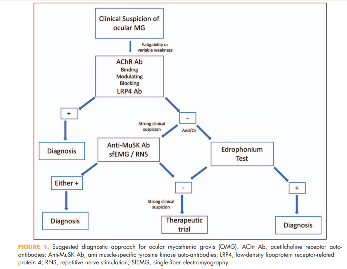
Antimuscle-specific tyrosine kinase and LDL-related receptor-related protein 4 are newly available serologictesting for myasthenia gravis that can help in increasing the diagnostic sensitivity of OMG. They should be included to the diagnostic algorithm of OMG in appropriate clinical situations.
Summary
OMG remains a primarily clinical diagnosis, but recent advances in laboratory testing can improve the diagnostic accuracy and should be used in appropriate clinical settings. The mainstay of treatment for OMG has not significantly changed over the past years, but the increasing availability of steroid-sparing agents improved the disease control while minimizing steroid-induced complications.

KEY POINTS
● OMG remains a clinical diagnosis, but various laboratory and electrophysiologic testing can help in increasing the diagnostic accuracy.
● Antimuscle-specific tyrosine kinase and LDL-related receptor-related protein 4 antibodies should be included in the diagnostic algorithm of patients suspected to have OMG.
● Therapy should aim at achieving satisfactory symptomatic control while reducing minimizing

Full Text https://drive.google.com/open?id=1ArU1gdYCwKWogLDCr0OcslzGd6egLN8y

Neuro-ophthalmology Illustrated Chapter 1 – Examination 5

Questions:
21. What are 5 clinical settings where OKN testing may be helpful?
22. Why should OKN testing be done in infants suspected of having the infantile nystagmus syndrome (congenital nystagmus)?
23. Where is the lesion likely to be located in a patient with homonymous hemianopia and symmetric OKN?
24. Where is the lesion likely to be located in a patient with homonymous hemianopia and asymmetric OKN response?

Neuro-ophthalmology Illustrated Chapter 1 – Examination 4

Questions:
17. Anisocoria more obvious in dim light indicates a sympathetic or parasympathetic lesion?
18. Dilation lag present when the lights are dimmed indicates a sympathetic or parasympathetic lesion?
19. How long after dimming the lights should one wait before checking for dilation lag?
20. Anisocoria more obvious in bright light indicates a sympathetic or parasympathetic lesion?

Neuro-ophthalmology Illustrated Chapter 1 – Examination 3

Questions:

11. Which 5 features of pupil function should be documented in a neuro-ophthalmic examination?

12. Would a Relative Afferent Pupillary Defect be expected with anisocoria?

13. Does an occipital lobe injury result in a Relative Afferent Pupillary Defect?

14. Does a unilateral optic neuropathy result in a Relative Afferent Pupillary Defect?

15. Can a unilateral optic tract lesion result in a Relative Afferent Pupillary Defect?

16. What anatomic factor explains the Relative Afferent Pupillary Defect with an optic tract lesion?

Recommended Reading – The expanding burden of idiopathic intracranial hypertension

Recommended Reading – The expanding burden of idiopathic intracranial hypertension

The expanding burden of idiopathic intracranial hypertension
Susan P. Mollan, Magda Aguiar, Felicity Evison, Emma Frew & Alexandra J. Sinclair. Eye (2018)

Free Full Text: https://www.nature.com/articles/s41433-018-0238-5

Abstract
OBJECTIVE: To quantify the hospital burden and health economic impact of idiopathic intracranial hypertension.

METHODS: Hospital Episode Statistics (HES) national data was extracted between 1st January 2002 and 31st December 2016. All those within England with a diagnosis of idiopathic intracranial hypertension were included. Those with secondary causes of raised intracranial pressure such as tumours, hydrocephalus and cerebral venous sinus thrombosis were excluded.

RESULTS: A total of 23,182 new IIH cases were diagnosed. Fifty-two percent resided in the most socially deprived areas (quintiles 1 and 2). Incidence rose between 2002 and 2016 from 2.3 to 4.7 per 100,000 in the general population. Peak incidence occurred in females aged 25 (15.2 per 100,000). 91.6% were treated medically, 7.6% had a cerebrospinal fluid diversion procedure, 0.7% underwent bariatric surgery and 0.1% had optic nerve sheath fenestration. Elective caesarean sections rates were significantly higher in IIH (16%) compared to the general population (9%), p < 0.005. Admission rates rose by 442% between 2002 and 2014, with 38% having repeated admissions in the year following diagnosis. Duration of hospital admission was 2.7 days (8.8 days for those having CSF diversion procedures). Costs rose from £9.2 to £50 million per annum over the study period with costs forecasts of £462 million per annum by 2030.

CONCLUSIONS: IIH incidence is rising (by greater than 100% over the study), highest in areas of social deprivation and mirroring obesity trends. Readmissions rates are high and growing yearly. The escalating population and financial burden of IIH has wide reaching implications for the health care system.

Composite figure.


a Incidence in the general population. b Incidence by age and gender. c Annual incidence in females and males and Obesity rates (% obesity per annum (body mass index ≥ 30), age-standardized in 18 years + by gender in the United Kingdom. From World Health organisation http://apps.who.int/gho/data/node.main.A900A?lang=en Accessed 6 Oct 2017. d Management of IIH in the cohort. e Geographical distribution of diagnosed cases of IIH in England. F Distribution of cases by region per annum.


Neuro-ophthalmology Illustrated Chapter 1 – Examination 2

Questions:
4. For the Photostress Recovery Test, how long does the patient look at a bright light held a few centimeters from the eye?
5 For the Photostress Recovery Test, what is the recovery end point to observe?
6. For the Photostress Recovery Test, what is the normal recovery time?
7. Which conditions prolong the recovery time of the Photostress Recovery Test?
8. What is the normal height of the palpebral fissure?
9. What is the normal Marginal Reflex Distance (MRD1)?
10. How many millimeters is the normal levator function measurement?