Archives for July 2018

Recommended Reading Cavernous Sinus Thrombosis

From:  MRI in the Evaluation of Acute Visual Syndromes.
Mukhi SV, Lincoln CM. Topics in Magnetic Resonance Imaging 24 (6):309-24. 2015
https://drive.google.com/open?id=1ayqCxDRn0E_Own4NkKoWbC2zqvfAc2X8

The prevalent use of antibiotics has decreased the overall incidence of CST. CST still carries significant mortality, commonly reported as approximately 30%, with more than 50% of the cases resulting in morbidity secondary to cranial neuropathies. CST is subclassified as aseptic or infectious in etiology. Aseptic causes include surgery or trauma. Infectious CST is typically a complication of a facial, orbital, odontogenic, or paranasal sinus infection. Sinusitis is the most common cause of CST, whereas odontogenic sources have been reported in up to 10% of the cases.3,33–39

The CS is a paired structure on either side of the sella, pituitary gland, and sphenoid sinus. It is composed of two layers of dura that are split to create a septate venous channel. The internal carotid artery (ICA) is the most medial structure and cranial nerves III, IV, and first and second branches of cranial nerve V are located in the lateral wall of the dura. Cranial nerve VI courses at the medial aspect of the ICA. Anteriorly, the CS is bordered by the SOF and OA. The posterior margin of the CS is immediately lateral to the dorsum sella and bordered by Meckel cave medially and the petrous apex posteroinferiorly.33,40,41

CST most commonly occurs secondary to the spread of infection by emissary veins as well as by direct extension. Emissary veins throughout the skull base are valve less and have bidirectional flow, accounting for the ease of contiguous spread.41 Spread of infection also occurs by the propagation of thrombus and/or septic embolism. It is postulated that bacteria stimulate the formation of thrombus by the release of a procoagulant substance and through toxins that cause tissue damage.38 In otitis media, infection spreads via the sigmoid sinus and along the internal carotid artery plexus. Staphylococcus aureus (70%) and Streptococcus sp (22%) are the important organisms responsible for infection of the CS. In patients with uncontrolled diabetes and immunocompromise, fungal infection can also be responsible, particularly mucormycosis.38,41

Tuberculosis has also been reported to cause both unilateral and bilateral CST; cavernous sinus tuberculoma may occur in the absence of pulmonary findings. Lymphomatous infiltration of the CS has been reported in both pediatric and adult patients.4

CST typically presents with orbital swelling, proptosis, chemosis, fever, and ophthalmoplegia. Visual impairment in CST has been reported in 7% to 22% of the cases, with blindness reported in 8% to 15% of the cases. As the disease progresses, decreased light perception and visual loss ensue. In a case report by Chen et al, CST-induced blindness suggested involvement of the bilateral retina and optic nerves. The postulated mechanisms accounting for visual impairment and blindness in CST include venous infarction of the retina and retinal ischemia caused by occlusion of either an ophthalmic artery branch or the central retinal artery, or by mechanical pressure at the OA.39

Chemosis, periorbital edema, and proptosis have been attributed to venous congestion.38 Papilledema as a result of raised intracranial pressure from a CST has been described as well.42 Palsies of III, IV, and VI cranial nerves secondary to compression result in impaired EOM motility. Intracranial extension of infection may result in meningitis, encephalitis, brain abscess, pituitary infection, epidural and subdural empyema, and coma/death.33,38,42

MRI is the radiologic examination of choice, and the CS should be imaged in its entirety. MRI demonstrates the contents of the CSs more effectively compared with CT.40 Imaging protocols should extend from the OA to the prepontine cistern. Routine T2, fluid-attenuated inversion recovery, and pre- and post-contrast T1 weighted images of the entire brain should be included. Postcontrast T1 weighted, 3-mm thick images should be obtained in the axial and coronal planes with at least one plane imaged utilizing a fat-saturation technique. Thin-section, postcontrast axial images may be acquired by three-dimensional spoiled gradient techniques. In addition, thin-section, three-dimensional, heavily T2 weighted images allow visualization of individual cranial nerves in the CS and adjacent cisterns.43 Pula et al describe the use of three-dimensional constructive interference in steady state to show smaller structures within the CS, making it the ideal choice to study cranial neuropathies in the CS.44

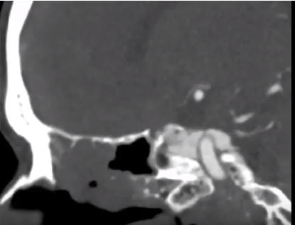
Alterations in signal intensity, size, and contour of the CS are subtle signs of thrombosis. A filling defect with enhancement of the peripheral margins of the CS suggests a clot within it (Fig. 4). Subacute thrombus exhibits high signal intensity on all pulse sequences, whereas acute thrombosis may appear more isointense. Indirect signs that may suggest the diagnosis are dilation of the superior ophthalmic veins, exophthalmos, and increased dural enhancement along the lateral border of CS and ipsilateral tentorium. Appropriate clinical symptoms, adjacent sinusitis, and orbital or odontogenic infection confirm the diagnosis and etiology.33,38,41,43

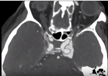
FIGURE 4. (A) Twenty-year-old man with invasive fungal sinusitis in setting of relapsed acute lymphocytic leukemia. Axial postcontrast image of the orbit and CS demonstrates filling defect in the left CS (arrow).
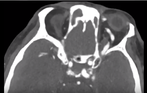
(B and C) Twenty-three-year old man with leukemia and rapidly progressive right-sided cranial neuropathy involving III, IV, and VI. Axial pre- (B) and post (C) contrast T1 images shows filling defect in the right CS with absence of the right cavernous carotid artery flow void (arrows).

CST therapy relies on mobilization of the varied disciplines of neurology, neurosurgery, otolaryngology, and infectious disease. Aggressive antibiotic therapy and surgical debridement of the primary site of infection and surrounding areas of involvement are the mainstay of treatment. The use of steroid therapy to reduce orbital edema and cranial nerve inflammation is controversial. Anticoagulant therapy has shown some benefit when initiated early.33,38,43

Neuro-ophthalmology questions of the week: NOI15-Cavernous Sinus and Orbital Vascular Disorders 3

Questions:
12. What are the findings of thrombosis of the cavernous sinus?
13. What is the usual cause of cavernous sinus thrombosis?
14. What should be considered in a patient with apparent orbital cellulitis?
15. What are the major risks of thrombosis of the cavernous sinus?
16. What are 3 very serious complications of cavernous sinus thrombosis?
17. What is the mnemonic for the structures and their position in the cavernous sinus?

Recommended Reading – Neuroimaging: Carotid Cavernous Fistula CT Angiogram Findings

CTisus  Published on Feb 10, 2017

These first five CTA images of the head demonstrate filling of the left cavernous sinus in the arterial phase and asymmetric enlargement and filling of the left superior ophthalmic vein.  These findings are consistent with a carotid cavernous fistula. The diagnostic angiogram was performed to evaluate the supply and drainage of the fistula. No aneurysm was identified. The fistula was supplied most prominently from the bilateral external carotid arteries and showed prominent retrograde drainage into the dilated left superior ophthalmic vein. These fistulas may present with unilateral or bilateral proptosis and chemosis and if severe may cause vision loss. This patient was treated with transvenous coil embolization and demonstrated no evidence of fistula on two month follow-up imaging.

Video https://www.youtube.com/watch?v=gkhd36dssqA

Reference
CT is us is created and maintained by The Advanced Medical Imaging Laboratory (AMIL). AMIL is a multidisciplinary team dedicated to research, education, and the advancement of patient care using medical imaging with a focus on spiral CT and 3D imaging. The AMIL is headed by Elliot K. Fishman, M.D.

http://www.ctisus.com


Neuro-ophthalmology questions of the week: NOI15-Cavernous Sinus and Orbital Vascular Disorders 2

Questions:
6. What are 10 ocular findings of carotid cavernous fistula?
7. Which of the cranial nerves is most commonly affected by a carotid cavernous fistula?
8. Can mechanical restriction of extraocular muscles occur in carotid cavernous fistula?
9. What diagnosis should be considered in an elderly woman with a mild headache, and elevated intraocular pressure?
10. What should be considered in all patients a bruit accompanying a chronically red eye?
11. When is treatment indicated for carotid cavernous fistulas?

_____________________________________

Neuro-ophthalmology questions of the week: NOI15-Cavernous Sinus and Orbital Vascular Disorders 1

Questions:
1. What signs and symptoms may develop from an aneurysm of the internal carotid artery within the cavernous sinus?
2. What are the characteristics of direct shunts between the internal carotid artery and the cavernous sinus?
3. What are the characteristics of indirect carotid cavernous or dural shunts?
4. Does a carotid cavernous fistula have unilateral or bilateral ocular symptoms?
5. Do carotid cavernous fistulas always have ocular symptoms?

Recommended Reading – Teaching NeuroImages: Ocular bruit in carotid-cavernous sinus fistula

Teaching NeuroImages: Ocular bruit in carotid-cavernous sinus fistula
Jeong-Yoon Choi, Seol-Hee Baek, Jin-Man Jung, Do-Young Kwon,
Moon Ho Park
Neurology. August 12, 2014; 83 (7) RESIDENT AND FELLOW SECTION

ARTICLE
A 57-year-old man who had a traffic accident 1 month previously presented with left ocular pain, double vision, and left eye proptosis with ptosis and conjunctival hemorrhage. Fundus showed dilated veins with no hemorrhages or disc edema. Left ocular motility showed complete external ophthalmoplegia (figure 1). There was prominent ocular bruit in his left eye (audio file on the Neurology® Web site at Neurology.org). MRI and magnetic resonance angiography showed a dilated left superior ophthalmic vein and an extravasation into cavernous sinus (figure 2). With chemosis, ophthalmoplegia, and retro-orbital pain, the auscultation of orbital bruit can make a correct and prompt diagnosis in the patient with carotid-cavernous sinus fistula.1


Figure 1 Physical examination
(A) Left eye with conjunctival injection and ptosis.

(B) Left eye proptosis.
(C) Fundus shows dilated veins with no hemorrhages or disc edema.
(D) Ocular motility shows complete external ophthalmoplegia in left eye and partial limitation of abduction in right eye.


Figure 2 Brain MRI and magnetic resonance angiography findings
(A) magnetic resonance angiography
(B) show a dilated left superior ophthalmic vein (black arrowhead) and a extravasation into cavernous sinus (white arrow).

Audio. Auscultation of ocular bruit.

(audio.mp3)
It was recorded using the JABES electronic stethoscope (GS tech., Korea) and WavePad Sound Editor (NCH software, Australia).

AUTHOR CONTRIBUTIONS Dr. Choi: participated in conceptualization of the manuscript, drafted the manuscript. Dr. Baek: participated in analysis of results and conceptualization of the manuscript. Dr. Jung: selected appropriate images and revised the manuscript for intellectual content. Dr. Kwon: participated in analysis of results and revised the manuscript for intellectual content. Dr. Park: drafted the manuscript and figure legend and revised the manuscript for intellectual content.

STUDY FUNDING
 
No targeted funding reported.
DISCLOSURE The authors report no disclosures relevant to the manuscript. Go to Neurology.org for full disclosures.
Footnotes
Go to Neurology.org for full disclosures. Funding information and disclosures deemed relevant by the authors, if any, are provided at the end of the article.
Supplemental data at Neurology.org
Download teaching slides: Neurology.org
© 2014 American Academy of Neurology

REFERENCE 1. Ling JD, Chao D, Al Zubidi N, Lee AG. Big red flags in neuro-ophthalmology. Can J Ophthalmol2013;48:3–7.


Neuro-ophthalmology questions of the week: NOI14 Orbital Syndrome

Questions:
1. What is the orbital syndrome?
2. What are the common features of the orbital syndrome?
3. What is the most common cause of unilateral or bilateral proptosis?
4. What should be suspected if there is globe displacement with proptosis?
5. Do brain CT and MRI often miss orbital processes?