Archives for June 2018

Teaching NeuroImages: The half-split man

Teaching NeuroImages: The half-split man
Makoto Takahashi, Akiko Shinya, Hisao Kitazono, Teruhiko Sekiguchi, Akira Inaba, Satoshi Orimo. Neurology. September 13, 2016; 87 (11) RESIDENT AND FELLOW SECTION

ARTICLE
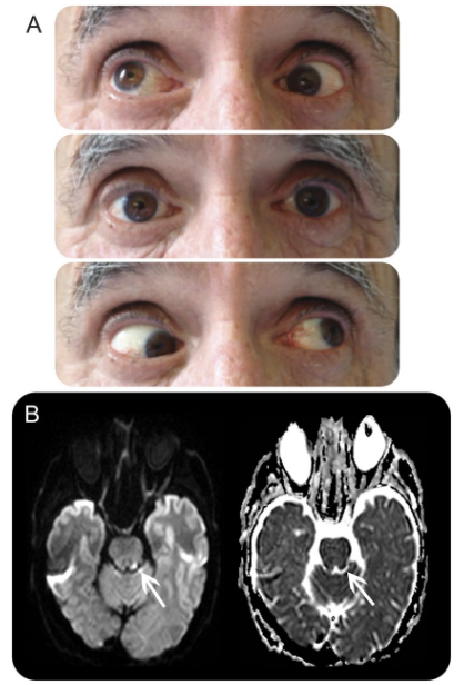
A 51-year-old man was admitted with left lateral medullary infarction due to vertebral artery dissection (figure 1). Neurologic examination revealed nystagmus, dissociated sensory disturbance, and no evidence of paralysis. Miosis and ptosis were observed on the ipsilateral side, but hypohidrosis was not apparent. Thermography revealed a bilateral discrepancy in body temperature, as if the patient were split down the middle (figure 2). Asymmetric skin temperature can occur among patients with Wallenberg syndrome associated with Horner syndrome due to a disturbance of the descending sympathetic tract that causes ipsilateral hypohidrosis and increased cutaneous blood flow.1

Figure 1 MRI and magnetic resonance angiography of the medulla and the vertebral artery
Diffusion-weighted and T2-weighted images show an acute infarction of the left lateral medulla (A, B). Magnetic resonance angiography and black-blood MRI show dissection of the left vertebral artery (C, D).

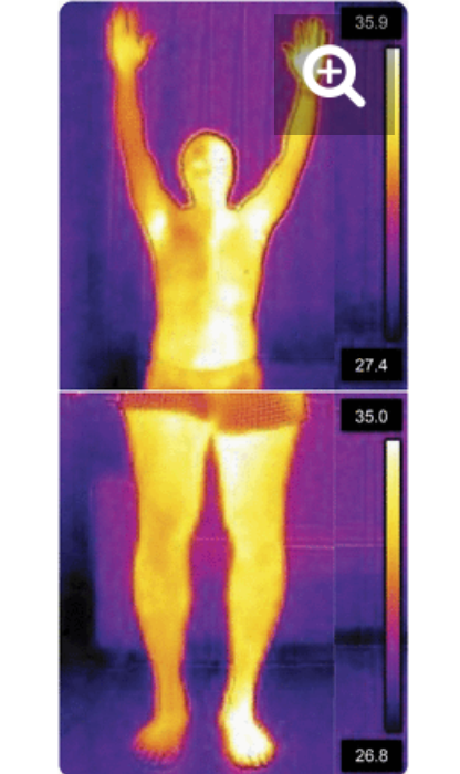
Figure 2 Thermography findings
Thermography images show the bilateral discrepancy in body temperature (in °C), as though the patient were split down the middle of his body.

AUTHOR CONTRIBUTIONS Dr. Takahashi: study concept, interpretation of data, and drafting the manuscript. Dr. Shinya: revision of the manuscript for intellectual content. Dr. Sekiguchi: study supervision. Dr. Kitazono: study supervision. Dr. Inaba: study supervision. Dr. Orimo: revision of the manuscript for intellectual content and study supervision.

STUDY FUNDING No targeted funding reported.

DISCLOSURE The authors report no disclosures relevant to the manuscript. Go to Neurology.org for full disclosures.

Footnotes Download teaching slides: Neurology.org

REFERENCE
1. Korpelainen JT, Sotaniemi KA, Myllylä VV. Asymmetrical skin temperature in ischemic stroke. Stroke 1995;26:1543–1547.


Neuro-ophthalmology Questions of the Week: NOI13 Diplopia10.3 – Other Eye Movement Abnormalities

Questions:
21. What is the Raymond syndrome?
22. What is the Millard-Gubler syndrome?
23. What is the Foville syndrome?
24. What is the Wallenberg syndrome?
25. What is the Weber syndrome?
26. What is the Nothnagel syndrome?
27. What is Benedikt syndrome?
28. What is the Claude syndrome?
29. What is the top of the basilar syndrome?
30. What systemic disorders can affect the ocular motor cranial nerves?

Teaching Video NeuroImages: Is it III alone, or III and IV?

Teaching Video NeuroImages: Is it III alone, or III and IV?
Stephen G. Reich

Neurology May 22, 2007; 68 (21)
RESIDENT AND FELLOW SECTION http://n.neurology.org/content/68/21/E34

Series editor: Mitchell S.V. Elkind MD, MS, Section Editor

The most important questions, when confronted with an oculomotor (III) palsy are:  
1) Is the pupil spared?
2) Is it complete aside from pupil sparing? and
3) Is it in isolation?

A “no” answer to any makes a benign, ischemic III palsy less likely.1

In the presence of a III palsy, the traditional method of testing the trochlear nerve (IV) at the bedside by asking the patient to depress the adducted eye cannot be performed. Instead, the patient should be instructed to abduct the eye and then look down; if IV is intact, there will be intorsion.2

Confirming that IV is intact in the presence of a III palsy is important because the combination of an oculomotor and trochlear palsy suggests a lesion in the cavernous sinus.

A 56-year-old man presented with a complete, pupil-sparing right oculomotor palsy (video E-1). The evaluation was negative, and the palsy resolved within 1 month.
Video
The video demonstrates a pupil-sparing but otherwise complete right oculomotor palsy.
There is ptosis. The eye is down, out, and unable to adduct, depress, or elevate. With attempted down gaze, there is intorsion, confirming that IV is intact. Although not demonstrated in the video, this primary action of IV should be tested by first having the patient abduct and then attempt to depress the eye. Intorsion is best appreciated by observing a medial conjunctival vessel.

ACKNOWLEDGMENT The author thanks Dr. Neil Miller for assistance.

Footnotes Disclosure: The author reports no conflicts of interest.

REFERENCES
1. Trobe JD. Isolated third nerve palsies. Sem Neurol 1986;6:135–141.
2. Ansons AM, Davis H. Diagnosis and management of ocular motility disorders. 3rd ed. Oxford: Blackwell Science Ltd, 2001:359–360.


Neuro-ophthalmology Questions of the Week: NOI13 Diplopia10.2 – Other Eye Movement Abnormalities

Questions:
11. What is the Tolosa-Hunt syndrome?
12. Where do these findings localize the lesion: Nystagmus, skew deviation, ocular tilt reaction, vertigo, lateropulsion, ipsilateral Horner syndrome, cerebellar syndrome, facial hypoesthesia, cranial nerves IX and X, and contralateral pain and thermal hypoesthesia (Wallenberg syndrome)?
13. Where do these findings localize the lesion: 4th nerve palsy with contralateral Horner syndrome?
14. Where do these findings localize the lesion: 3rd nerve palsy with contralateral ptosis and contralateral superior rectus weakness?
15. Where do these findings localize the lesion: 3rd nerve palsy with contralateral hemiparesis (Weber syndrome)?
16. Where do these findings localize the lesion: 3rd nerve palsy and ipsilateral cerebellar ataxia (Nothnagel syndrome)?
17. Where do these findings localize the lesion: 3rd nerve palsy and contralateral tremor (Benedikt syndrome)?
18. Where do these findings localize the lesion: 3rd nerve palsy and contralateral ataxia with tremor (Claude syndrome)?
19. Where do these findings localize the lesion: 3rd nerve palsy with vertical gaze palsy, lid retraction, skew deviation, and convergence nystagmus?
20. Where do these findings localize the lesion: 3rd nerve palsy with depressed mental status?  

Pearls and oy-sters of localization in ophthalmoparesis

Pearls and oy-sters of localization in ophthalmoparesis
Teresa Buracchio, Janet C. Rucker
Neurology. December 11, 2007; 69 (24) RESIDENT AND FELLOW SECTION

Abstract
Ocular misalignment and ophthalmoparesis result in the symptom of binocular diplopia. In the evaluation of diplopia, localization of the ocular motility disorder is the main objective. This requires a systematic approach and knowledge of the ocular motor pathways and actions of the extraocular muscles. This article reviews the components of the ocular motor pathway and presents helpful tools for localization and common sources of error in the assessment of ophthalmoparesis.

Neuro-ophthalmology Questions of the Week: NOI13 Diplopia10.1 – Other Eye Movement Abnormalities

Questions:
1. What are the findings of the Locked-in Syndrome?
2. Where is the lesion in the Locked-in Syndrome?
3. What is Ocular Neuromyotonia?
4. What are the symptoms of Ocular Neuromyotonia?
5. What is the usual cause of Ocular Neuromyotonia?
6. Where do these findings localize the lesion: Horizontal gaze palsy with ipsilateral facial palsy?
7. Where do these findings localize the lesion: 6th nerve palsy with contralateral hemiparesis (Raymond syndrome)?
8. Where do these findings localize the lesion: 6th nerve palsy with ipsilateral seventh nerve palsy and contralateral hemiparesis (Millard-Gubler syndrome)?
9. Where do these findings localize the lesion: 6th nerve palsy with ipsilateral seventh nerve palsy, deafness, hypoesthesia, Horner syndrome, contralateral pain and thermal hypoesthesia, ataxia (Foville syndrome)?
10. Where do these findings localize the lesion: 6th nerve palsy with ipsilateral Horner Syndrome?

Teaching Video NeuroImages: Alternating skew deviation with abducting hypertropia following superior colliculus infarction

Teaching Video NeuroImages: Alternating skew deviation with abducting hypertropia following superior colliculus infarction
Damien Biotti, Marianne Barbieux and David Brassat
Neurology. March 01, 2016; 86 (9) RESIDENT AND FELLOW SECTION

A 63-year-old patient was admitted with acute ataxia and binocular oblique diplopia. Neuro-ophthalmologic examination revealed abducting hypertropia on lateral gaze, better seen during upgaze, mimicking bilateral inferior oblique palsy (Figure A, video). There was no ocular cyclotorsion. Brain MRI revealed focal ischemic lesions in the right cerebellar hemisphere and left superior colliculus (Figure B). The diagnosis of alternating abducting hypertrophic skew deviation was made. This rare type of skew deviation is related to central otolithic dysfunction. Similar cases have been described with cerebellar, pretectal, or cervico-medullary junction lesions.1,2 Orthoptic management can help and patients can slowly improve over months.


Figure  Ocular motor examination and MR images
(A) Ocular motor examination (right gaze, straight gaze, left gaze).
(B) Diffusion-weighted imaging (left) and apparent diffusion coefficient images (right) reveal a focal and acute ischemic stroke.

AUTHOR CONTRIBUTIONS D. Biotti: principal author, corresponding author. M. Barbieux: contributor, neurologic management. D. Brassat: contributor.

STUDY FUNDING No targeted funding reported.

DISCLOSURE The authors report no disclosures relevant to the manuscript. Go to Neurology.org for full disclosures.

Footnotes
  ● Go to Neurology.org for full disclosures. Funding information and disclosures deemed relevant by the authors, if any, are provided at the end of the article.
● 
Supplemental data at Neurology.org
  ● Download teaching slides: Neurology.org

REFERENCES
1. Versino M, Hurko O, Zee D. Disorders of binocular control of eye movements in patients with cerebellar dysfunction. Brain 1996;119:1933–1950.
2. Hamed LM, Maria BL, Quisling RG, Mickle JP. Alternating skew on lateral gaze: neuroanatomic pathway and relationship to superior oblique overaction. Ophthalmology 1993;100:281–286.


Neuro-ophthalmology questions of the week: NOI13 Diplopia 9.4 – Internuclear & Supranuclear Lesions

Questions:
38. Where are eye movements initiated?
39. Which of the following can cause a patient to be unable look to the left?
      A. Right FEF, B. Left FEF, C. Right PPRF, D. Left PPRF
40. Where are smooth pursuit eye movements generated?
41. What is the function of inputs to the visual system from the vestibular nuclear complexes?
42. What is the cause of skew deviation?