Recommended Reading – Unusual presentation of an Adie-like pupil Cat’s eye pupil

Unusual presentation of an Adie-like pupil Cat’s eye pupil
Robert A. Egan, Carla Avruskin
NEUROIMAGES
Neurology. 2018; 91 (15)

Article
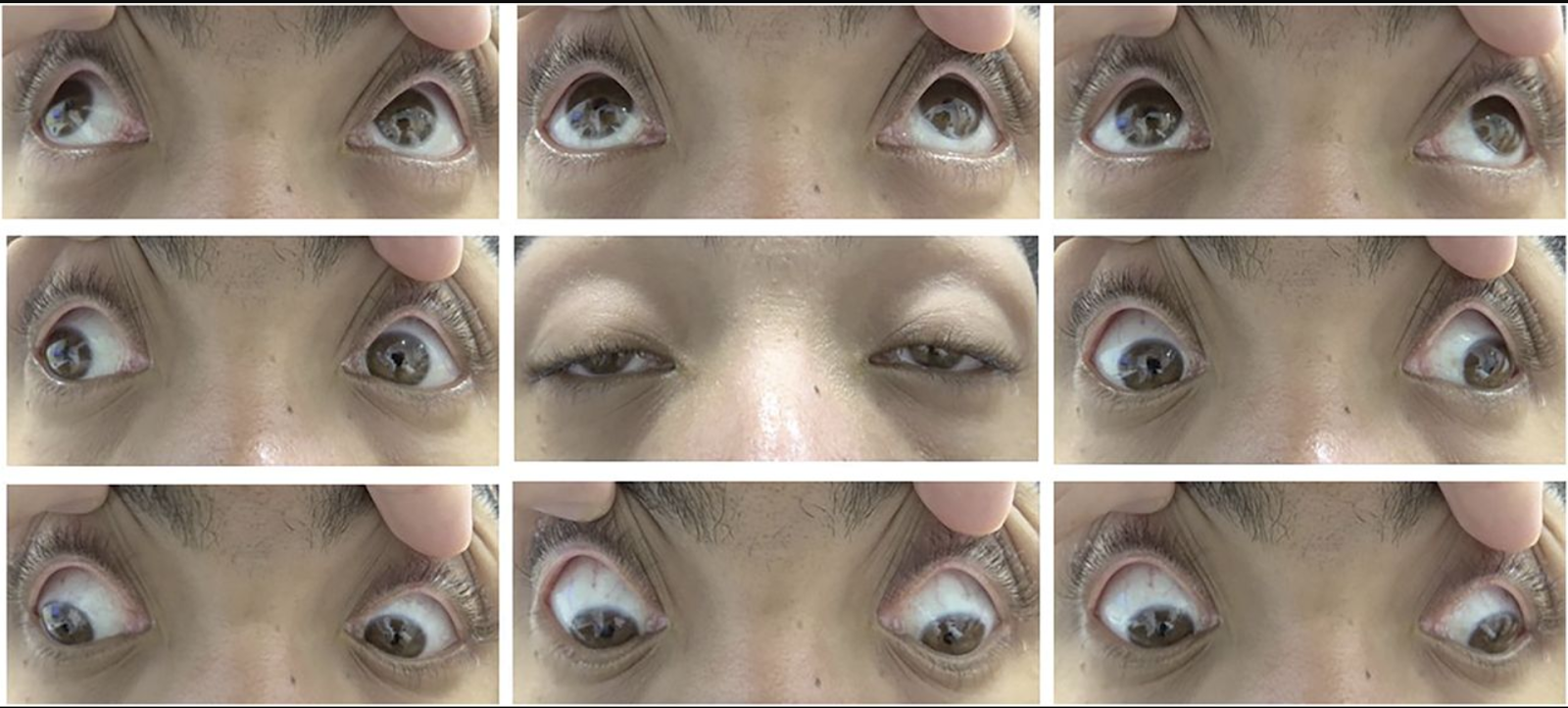
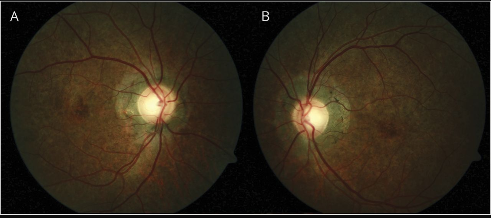
A 55-year-old woman noticed that her right pupil was oddly shaped, associated with a mild ache. Her afferent visual examination, ocular and eyelid motility, and fundi were normal. Her left pupil was round and reactive (figure 1).

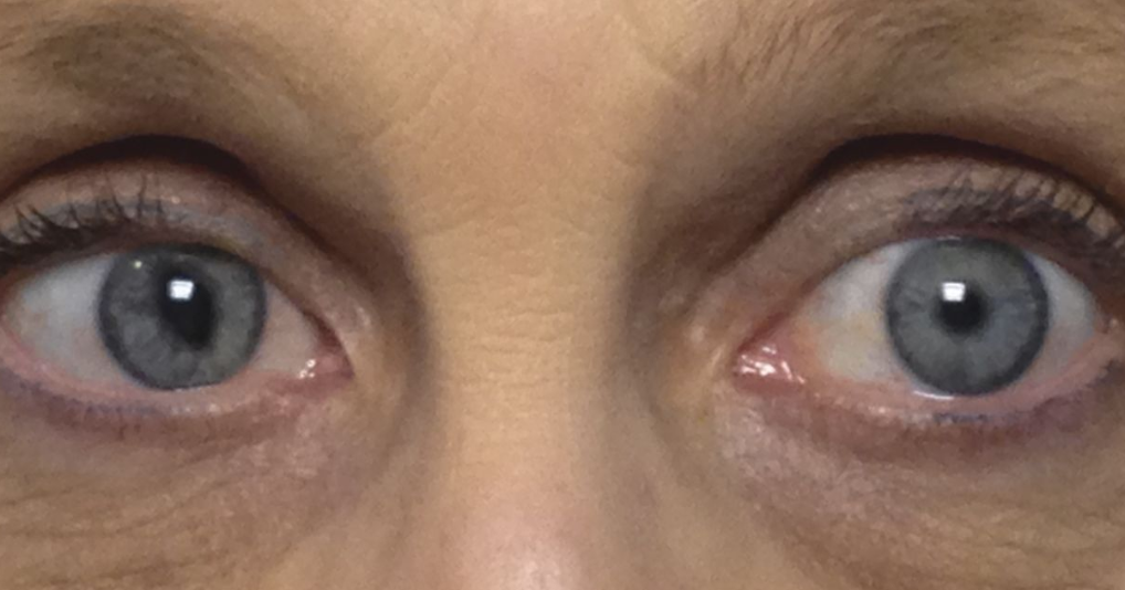
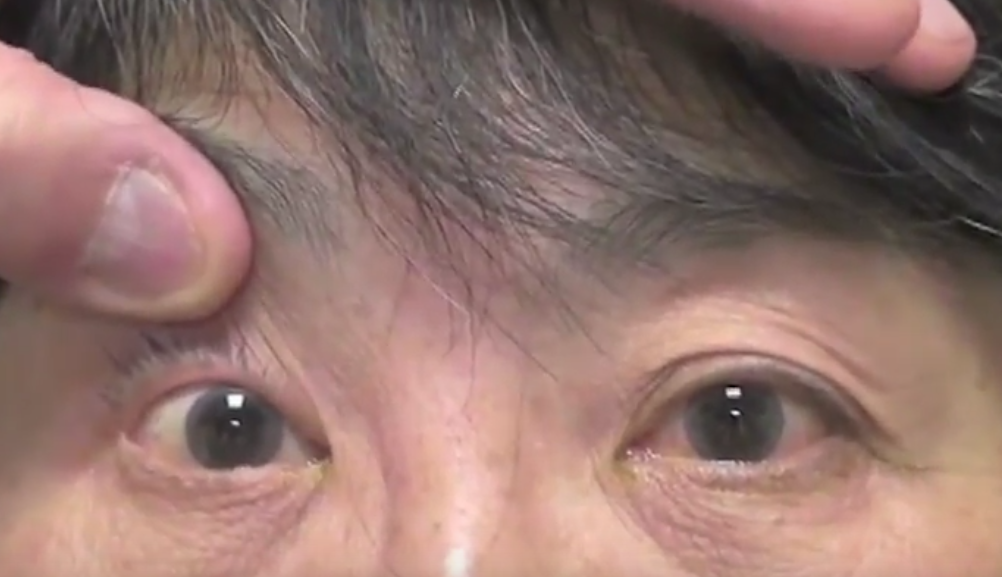
Figure 1 Close-up shows normal left pupil and tonic right cat’s eye pupil
The right pupil was elongated (points at 11:00 and 5:00) and tonic (figure 2).

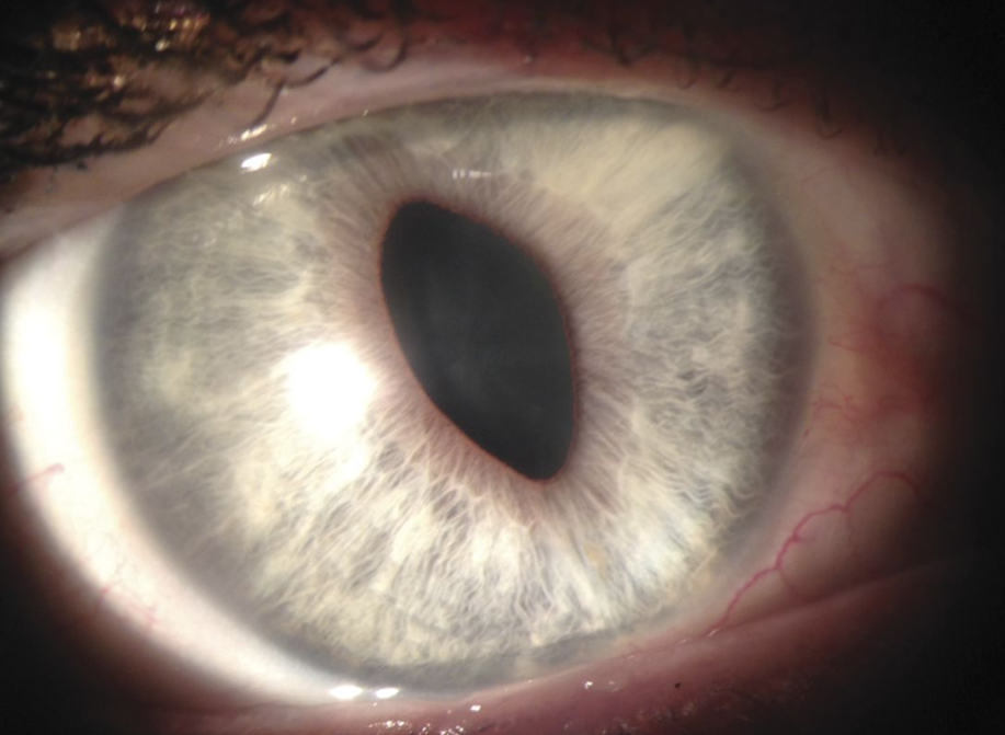
Figure 2 There was sectoral paralysis of the iris between 12:00 and 4:00 and also from 6:00 to 10:00 with mild thinning of the pupillary ruff on the temporal iris characteristic of an Adie-like pupil

There was no ocular hypertension or corneal edema. A year later, the pupil shape became triangular; evaluation revealed ocular hypertension without corneal edema, confirming the iridocorneal endothelial (ICE) syndrome. This case is an unusual presentation of a cat’s eye Adie-like pupil as the harbinger for ICE syndrome.

Recommended Reading – Teaching NeuroImages: Kearns-Sayre syndrome

Teaching NeuroImages: Kearns-Sayre syndrome 
Michael T.B. Nguyen, Jonathan Micieli, Edward Margolin
RESIDENT & FELLOW SECTION. Neurology. 2019; 92 (5)

Article

A 19-year-old man presented 6 months postimplantation of permanent pacemaker for complete heart block with bilateral nonfatigable symmetric ptosis, diminished levator superioris function, and symmetric ophthalmoplegia (figure 1). Funduscopy revealed bilateral pigmentary retinopathy (figure 2). Skeletal muscle biopsy revealed presence of ragged-red fibers, consistent with Kearns-Sayre syndrome. This mitochondrial disorder is characterized by the triad of onset before age 20, chronic progressive external ophthalmoplegia, and pigmentary retinopathy. Other findings can include complete heart block, cerebellar ataxia, deafness, and endocrinopathies. CSF folate levels should be measured and supplemented if low. There is no definitive treatment but annual surveillance for comorbidities is required.1,2

Figure 1 Ptosis and ophthalmoplegia
Photographs of eye movement demonstrate severe bilateral ptosis and mild diffuse ophthalmoplegia in all directions of attempted gaze.

Figure 2 Pigmentary retinopathy
Fundus examination of right (A) and left eye (B) shows bilateral pigmentary retinopathy. There is diffuse depigmentation of the retinal pigment epithelium in a salt-and-pepper pattern of pigment clumping and involvement of the peripapillary zone.

References
1. Kearns TP, Sayre GP. Retinitis pigmentosa, external ophthalmoplegia, and complete heart block. Arch Ophthalmol 1958;60:280–289.
2. Shemesh A, Margolin E. Kearns Sayre syndrome. In: StatPearls [Internet]. Treasure Island, FL: StatPearls Publishing; 2018.

Recommended Reading – Volitional opsoclonus

Recommended Reading – Volitional opsoclonus


Volitional opsoclonus
.

Patricio S. Espinosa, Joseph R. Berger. Neurology. 2005; 65 (5)

NEUROIMAGES


Opsoclonus (saccadomania) is defined as random, uncalled-for, large-amplitude, back-to-back saccades without intersaccadic intervals. An abnormality of the omnipause neurons in the brainstem nucleus raphe interpositus has been suggested, but not proven, as the etiology of opsoclonus.
1 Opsoclonus occurs with paraneoplastic, infectious, postinfectious, toxic, or metabolic disorders; however, it is often idiopathic. Intentional fixation instability may result in ocular movements closely resembling opsoclonus.2 We describe a patient with the volitional ability to replicate “opsoclonus.”


This 55-year-old man presented with transient left-sided numbness which, after thorough evaluation, was attributed to a transient ischemic episode. His neurologic examination, including ocular motility, was perfectly normal. However, he was voluntarily able to display wildly chaotic eye movements (video clip). He commented that he has been aware of this ability since childhood. No other family members, to the best of his knowledge, are able to replicate these eye movements. Analysis of the eye movements permitted by looping and pausing the video segment reveals that the actual eye movements are either diagonal (horizontal and vertical in phase) or, in some cases, elliptical (horizontal and vertical 90º out of phase), indicating that it is “voluntary opsoclonus,” also referred to as “voluntary multiplanar flutter” (Louis Dell’Osso, Cleveland, OH, personal communication, January 4, 2005).


The identification of volitional eye movements in clinical practice is important because it permits the distinction between neurologic and factitious disorders.

Video https://drive.google.com/open?id=1_mHS61kcc5YPUpPL_VeU5EYTVtaUoYE_

References:


1. Ridley A, Kennard C, Scholtz CL, Buttner-Ennever JA, Summers B, Turnbull A. Omnipause neurons in two cases of opsoclonus associated with oat cell carcinoma of the lung. Brain 1987;110(Pt 6):1699–1709.

2. Yee RD, Spiegel PH, Yamada T, Abel LA, Suzuki DA, Zee DS. Voluntary saccadic oscillations, resembling ocular flutter and opsoclonus. J Neuroophthalmol 1994;14:95–101.


Recommended Reading – Pupillary sign of aberrant regeneration of the third nerve

Recommended Reading – Pupillary sign of aberrant regeneration of the third nerve

Pupillary sign of aberrant regeneration of the third nerve
Olga R. Rosenvald, Simmons Lessell. Neurology. 2016; 86 (18)
VIDEO NEUROIMAGES

A 55-year-old woman presented with a third nerve palsy and impaired abduction of her right eye. MRI showed a lesion at the orbital apex extending into the cavernous sinus. A biopsy showed invasive Aspergillus fumigatus and she was treated with antifungals with only partial improvement.

Twelve years later, she had complete ptosis, partially restricted abduction, adduction, and depression of the right eye. The right pupil, which did not constrict to light or near vision, constricted on downgaze (video on the Neurology® Web site at Neurology.org), diagnostic of aberrant regeneration,1 which occurs when regenerating axons are misdirected to muscles they were not intended, such as the iris sphincter.2

Video  https://drive.google.com/open?id=1D0qDczD0jsQ7q88eUL0KzVmMhlIOqqXf

The video shows a partial cranial nerve III palsy with a pupil reactive to neither light or near vision. However, the pupil constricts on attempted downgaze.

References:
1. Czarnecki JS, Thompson HS. The iris sphincter in aberrant regeneration of the third nerve. Arch Ophthalmol 1978;96:1606–1610.
2. Sibony PA, Lessell S, Gittinger JW. Acquired oculomotor synkinesis. Surv Ophthalmol 1984;28:382–390.


Recommended Reading – Incidence and Causes of Overdiagnosis of Optic Neuritis.

Incidence and Causes of Overdiagnosis of Optic Neuritis.
Stunkel L, Kung NH, Wilson B, McClelland CM, Van Stavern GP.
JAMA Ophthalmol. 2018 Jan 1;136(1):76-81.  

Abstract
IMPORTANCE: Diagnostic error is an important source of medical error. Overdiagnosis of optic neuritis may prompt unnecessary and costly diagnostic tests, procedures, and treatments.

OBJECTIVE: To assess the incidence of and characterize factors contributing to overdiagnosis of acute optic neuritis.

DESIGN, SETTING, AND PARTICIPANTS: In this retrospective clinic-based cross-sectional study of new patient encounters, 122 patients referred for acute optic neuritis at a university-based Midwestern neuro-ophthalmology clinic between January 2014 and October 2016 were studied. Data were analyzed from September 2016 to July 2017.

INTERVENTIONS: Definite diagnosis was determined by neuro-ophthalmologists. For patients with alterative diagnoses, the Diagnosis Error Evaluation and Research taxonomy tool was applied to categorize the type of diagnostic error.

MAIN OUTCOMES AND MEASURES: The primary outcome was the primary type of diagnostic error in patients erroneously diagnosed as having optic neuritis. Secondary outcomes included final diagnosis and interventions undergone prior to referral.

RESULTS: A total of 122 patients were referred with acute optic neuritis during the study period; 88 (72.1%) were female, and the mean (SD) age was 42.6 (14.0) years. Of these, 49 patients (40.2%; 95% CI, 31.4-49.4) were confirmed to have optic neuritis, and 73 (59.8%; 95% CI, 50.6-68.6) had an alternative diagnosis. The most common alternative diagnoses were headache and eye pain, functional visual loss, and other optic neuropathies, particularly nonarteritic anterior ischemic optic neuropathy. The most common diagnostic error was eliciting or interpreting critical elements of history, which occurred in 24 of 73 patients (33%) with alternative diagnoses. Other common errors included errors weighing or considering alternative diagnoses (23 patients [32%]), errors weighing or interpreting physical examination findings (15 patients [21%]), and misinterpreting diagnostic test results (11 patients [15%]). In patients with alternative diagnoses, 12 (16%) had normal magnetic resonance imaging findings preceding the referral, 12 (16%) had received a lumbar puncture, and 8 (11%) had received unnecessary treatment with intravenous steroids.

CONCLUSIONS AND RELEVANCE: These data suggest that nearly 60% (95% CI, 50.6-68.6) of patients referred for optic neuritis have an alternative diagnosis, with the most common errors being over reliance on a single item of history and failure to consider alternative diagnoses. Understanding pitfalls leading to overdiagnosis of optic neuritis may improve clinicians’ diagnostic process.

Free Full Text: https://www.ncbi.nlm.nih.gov/pmc/articles/PMC5833602/

___________________________________________________

Medscape Commentary on Above Article
https://www.medscape.com/viewarticle/907024
Errors Abound in Diagnosing Optic Neuritis–How Can We Fix Them?
Brianne N. Hobbs, OD. January 03, 2019

Thinking about mistakes, especially medical mistakes, is uncomfortable. It is certainly more enjoyable to read about a promising new therapy than to scrutinize the reasons that medical mistakes are happening. Yet, medical mistakes are currently the third leading cause of death in the United States, and despite their prevalence, there is no systematic way of reporting such errors.[1]

Death is not the only adverse outcome of medical errors, and it is estimated that mistakes causing serious harm are 10-20 times more common than those causing lethal harm.[2] Misdiagnosis is one category of medical mistakes that may result in unnecessary referrals, procedures, and treatments. A seemingly simple way to deliver better care is to identify common mistakes and correct them. This process isn’t glamorous, but it is productive.

Focus on Optic Neuritis

In a recent study,[3] one large university-based neuro-ophthalmology clinic addressed the problem of misdiagnosis of optic neuritis. Over a 2-year period, the clinic received 122 referrals for optic neuritis. The records for each of these patients were examined retrospectively to determine whether the diagnosis of optic neuritis was accurate and, if not, the reasons that commonly led to misdiagnosis.

Alarmingly, 73 of the 122 patients referred to this center with a diagnosis of optic neuritis were actually misdiagnosed. This misdiagnosis rate of approximately 60% was consistent among all referral sources, including optometrists, ophthalmologists, and neurologists. The most common alternative diagnoses were headache with concurrent eye pain (22%), functional vision loss (19%), and other optic neuropathies (16%), including nonarteritic ischemic optic neuropathy (NAION; 12%). Patients older than 50 years were more likely to be misdiagnosed with optic neuritis.

The Path to Misdiagnosis

Why were so many patients misdiagnosed as having optic neuritis? The most common error was failure to interpret the critical elements of the history, which occurred in 33% of the patients. Specifically, an overreliance on a previous diagnosis of multiple sclerosis or optic neuritis led to inaccuracy in diagnosis. Additionally, too much emphasis was often given to eye pain or pain with eye movement, which was found in the referral notes of 29% of patients ultimately diagnosed with a different condition. Another source of diagnostic error was ignoring normal exam findings; the possibility of optic neuritis can almost be excluded entirely if the complete ocular examination is normal. Red cap desaturation testing was often given too much emphasis, resulting in the diagnosis of optic neuritis solely on one abnormal, subjective test.

Although the misdiagnosis of optic neuritis is not a lethal mistake, multiple patients received expensive and invasive procedures, such as MRI and lumbar punctures, which were not indicated. Eight patients were treated unnecessarily with IV steroids. Four patients diagnosed with optic neuritis actually had neoplasms. The cost of misdiagnosis was not death, but it did result in patient harm.

There are five major clinical applications of this study:
1. Optic neuritis almost always results in an abnormal MRI due to T2 hyperintensity or enhancement of the optic nerve; a normal MRI argues strongly against optic neuritis.
2. The presence of an afferent pupillary defect was strongly correlated with a true diagnosis of optic neuritis; only 21% of patients who had been documented as previously having normal pupillary testing were ultimately diagnosed with optic neuritis.
3. Pain upon eye movement is often caused by a headache with associated eye pain or visual disturbances and is not pathognomonic for optic neuritis.
4. NAION is often misdiagnosed as optic neuritis in elderly patients, even though NAION has a higher incidence in this demographic.
5. No single element of the case history should determine the diagnosis, as this leads to diagnosis bias and a failure to consider other possible diagnoses.

Atul Gawande, in his book Complications: A Surgeon’s Notes on an Imperfect Science,[4] writes that “not only do all human beings err, but they err frequently and in predictable patterned ways.” Gathering data about these “predictable patterned ways” that clinicians err would help describe the scope of the problem, identify specific cognitive biases that lead to misdiagnosis, and generate explicit recommendations to improve patient care.

While this study was small and limited in scope, the methodology has broad applicability in enhancing patient care by reducing medical mistakes.

References:
1. Makary MA, Daniel M. Medical error-the third leading cause of death in the US. BMJ. 2016;353:i2139.
2. James JT. A new, evidence-based estimate of patient harms associated with hospital care. J Patient Saf. 2013;9:122-128.
3. Stunkel L, Kung NH, Van Stavern GP. Incidence and causes of overdiagnosis of optic neuritis: physician insecurity-reply. JAMA Ophthalmol. 2018;136:1312-1313.
4. Gawande A. Complications: A Surgeon’s Notes on an Imperfect Science. New York, NY: Picador; 2002.

Recommended Reading – Optical coherence tomography is a useful tool in the differentiation between true edema and pseudoedema of the optic disc.

Recommended Reading – Optical coherence tomography is a useful tool in the differentiation between true edema and pseudoedema of the optic disc.

Optical coherence tomography is a useful tool in the differentiation between true edema and pseudoedema of the optic disc.
PLoS One. 2018 Nov 29;13(11):e0208145. Carta A, Mora P, Aldigeri R, Gozzi F, Favilla S, Tedesco S, Calzetti G, Farci R, Barboni P, Bianchi-Marzoli S, Fossarello M, Gandolfi S1, Sadun AA.

Abstract
PURPOSE: To assess the usefulness of spectral-domain optical coherence tomography (SD-OCT) peripapillary retinal nerve fiber layer (RNFL) thickness measurement in discriminating early phase optic disc edema (ODE) from pseudoedema (PODE).

METHODS: Hospital-based, multicenter, cross-sectional study involving external patients referred for recent identification of “presumed ODE”. Patients underwent SD-OCT optic nerve head (ONH) RNFL thickness measurement at their first evaluation. In 155 of these, the causative etiology was subsequently ascertained and the respective eyes (one per patient) were assigned to the ODE (95 eyes) or PODE (60 eyes) group. Admission SD-OCT data were retrieved and used for the analysis. ROC curve analysis was used to calculate specificity, sensitivity and predictive value (PV) of the RNFL values.

RESULTS: The PODE group was significantly younger than the ODE group (p = 0.007). The average and any single-quadrant RNFL thickness was significantly higher in the ODE group compared with the PODE and control groups. The average and the inferior quadrant thicknesses tested the most powerful parameters to differentiate ODE from PODE. A cutoff value of ≥ 110 μm for the average area, or of ≥ 150 μm for the inferior quadrant was associated with maximal sensitivity and specificity with positive PV greater than 80%.

CONCLUSIONS: The SD-OCT evaluation of the peripapillary RNFL achieved good specificity, sensitivity and positive PV in discriminating between ODE and PODE. Despite the correct differential diagnosis between these categories still relies on a careful medical history taking and other ancillary testing, we proved the usefulness of SD-OCT RNFL measurement in supporting the diagnostic process.

Free Full Text https://www.ncbi.nlm.nih.gov/pmc/articles/PMC6264818/

Recommended Reading – Diagnostic Errors in Initial Misdiagnosis of Optic Nerve Sheath Meningiomas

Recommended Reading – Diagnostic Errors in Initial Misdiagnosis of Optic Nerve Sheath Meningiomas

Diagnostic Errors in Initial Misdiagnosis of Optic Nerve Sheath Meningiomas
Pinar Kahraman-Koytak, MD; Beau B. Bruce, MD, PhD; Jason H. Peragallo, MD; Nancy J. Newman, MD; Valérie Biousse, MD.  JAMA Neurol. Published online December 17, 2018.

IMPORTANCE Diagnostic errors can lead to the initial misdiagnosis of optic nerve sheath meningiomas (ONSM), which can lead to vision loss.

OBJECTIVE To identify factors contributing to the initial misdiagnosis of ONSM.

DESIGN, SETTING, AND PARTICIPANTS We retrospectively reviewed 35 of 39 patients with unilateral ONSM (89.7%) who were seen in the tertiary neuro-ophthalmology practice at Emory University School of Medicine between January 2002 and March 2017. The Diagnosis Error Evaluation and Research taxonomy tool was applied to cases with missed/delayed Diagnoses.

EXPOSURES Evaluation in a neuro-ophthalmology clinic.

MAIN OUTCOMES AND MEASURES Identifying the cause of diagnostic errors for patients who initially received a misdiagnosis who were found to have ONSM.

RESULTS Of 35 patients with unilateral ONSM (30 women [85.7%]; mean [SD] age, 45.26 [15.73] years), 25 (71%) had a diagnosis delayed for a mean (SD) of 62.60 (89.26) months. The most common diagnostic error (19 of 25 [76%]) was clinician assessment failure (errors in hypothesis generation and weighing), followed by errors in diagnostic testing (15 of 25 [60%]). The most common initial misdiagnosis was optic neuritis (12 of 25 [48%]), followed by the failure to recognize optic neuropathy in patients with ocular disorders. Five patients who received a misdiagnosis (20%) underwent unnecessary lumbar puncture, 12 patients (48%) unnecessary laboratory tests, and 6 patients (24%) unnecessary steroid treatment. Among the 16 patients who initially received a misdiagnosis that was later correctly diagnosed at our institution, 11 (68.8%) had prior magnetic resonance imaging (MRI) results that were read as healthy; 5 (45.5%) showed ONSM but were misread by a non-neuroradiologist and 6 (54.5%) were performed incorrectly (no orbital sequence or contrast). Sixteen of the 25 patients (64%) had a poor visual outcome. 

CONCLUSIONS AND RELEVANCE Biased preestablished diagnoses, inaccurate funduscopic examinations, a failure to order the correct test (MRI brain/orbits with contrast), and a failure to correctly interpret MRI results were the most common sources of diagnostic errors and delayed diagnosis with worse visual outcomes and increased cost (more visits and tests). Easier access to neuro-ophthalmologists, improved diagnostic strategies, and education regarding neuroimaging should help prevent diagnostic errors.

Full Text: https://drive.google.com/open?id=1sZB52iNgoN7LdAkT0rMDeB6tc6WsL9zM


Recommended Reading – Neuro-ophthalmological manifestations of Behçet’s disease

Recommended Reading – Neuro-ophthalmological manifestations of Behçet’s disease

Neuro-ophthalmological manifestations of Behçet’s disease
Ammr Alghamdi, Bahram Bodaghi, Chloé Comarmond, Anne Claire Desbois, Fanny Domont, Bertrand Wechsler, Raphael Depaz, Phuc Le Hoang, Patrice Cacoub, Valérie Touitou, David Saadoun
British Journal of Ophthalmology. 2018

Abstract
Background The neuro-ophthalmological manifestations of Behcet’s disease (BD) are rare, and data regarding their characteristics and outcome are lacking.

Objective To report prevalence, main characteristics and outcome of neuro-ophthalmological manifestations in BD.

Patients and methods This is a retrospective monocentric study of 217 patients diagnosed with neuro-Behçet’s disease (NBD), of whom 29 (13.3%) patients presented with neuro-ophthalmological manifestations (55% of men and mean±SD age of 26±8 years). All patients underwent a detailed ophthalmological examination and were followed up in the internal medicine and the ophthalmology departments.

Results Neuro-ophthalmological manifestations were the first presentation of BD in 45% of patients and developed later in the course of the disease in 55% of patients. They are divided into parenchymal (PM) and non-parenchymal (NPM)-related manifestations in, respectively, 13 patients (45%) and 16 patients (55%). PM included papillitis in seven patients (53.8%), retrobulbar optic neuritis in four patients (30.8%) and third cranial nerve palsy in two patients (15.4%). NPM included papilloedema related to cerebral venous thrombosis in all 16 patients, of whom 6 patients (37.5%) had sixth cranial nerve palsy. At initial examination, 93.1% of patients had visual alterations, including mainly decreased visual acuity visual field defects and/or diplopia. All patients were treated with corticosteroids and 79% of patients received immunosuppressive agents. After treatment, the visual outcome improved or stabilised in 66.7% of patients while it worsened in 33.3. The mean±SD logarithm of the minimum angle of resolution visual acuity improved from 0.4±0.3 at diagnosis to 0.2±0.3 after therapy. 10.3% and 3.4% of patients were legally blind at diagnosis and after therapy, respectively.

Conclusion Neuro-ophthalmological manifestations of BD represented 13% of NBD. They could be potentially severe and disabling. Prompt treatment is the key factor in improving visual outcome.


Full Text https://drive.google.com/file/d/18HvWBld3APQQR0WC4pp4zBtErv78G2-N/view?usp=sharing  

Recommended Reading – Teaching Video NeuroImages: Oculopalatal myoclonus

Recommended Reading – Teaching Video NeuroImages: Oculopalatal myoclonus

Teaching Video NeuroImages: Oculopalatal myoclonus
A possible consequence of brainstem injury
Neurology. November 27, 2018; 91 (22) RESIDENT & FELLOW SECTION

A 61-year-old man with previous left cerebellar infarct complained of double vision and dizziness several months postinfarct. Infarct is shown in the figure. Examination showed rotatory nystagmus in all directions of gaze, worse on left gaze (video 1). Oral examination showed palatal tremor (video 2). Oculopalatal tremor (OPT) is thought to result from interruption of connections between the red nucleus (midbrain), dentate nucleus (cerebellum), and inferior olive (medulla).1,2 The most common cause is brainstem infarction or hemorrhage,1 but it has also been observed with multiple sclerosis and other inflammatory entities. OPT usually becomes apparent several months after the injury. Treatment may include gabapentin, memantine, benzodiazepines, and valproic acid.

Figure: Brain MRI, fluid-attenuated inversion recovery
Image shows the patient’s infarct, involving the caudal portion of the left cerebellar hemisphere. Area of involvement corresponds to posterior inferior cerebellar artery territory.

Video 1: Rotatory nystagmus toward the left in all directions of gaze but worse on left and upward gaze. Rotatory nystagmus improved in amplitude and frequency with low dose clonazepam. http://movie-usa.glencoesoftware.com/video/10.1212/WNL.0000000000006563/video-1

Video 2: Video shows rhythmic contractions of the soft palate.
http://movie-usa.glencoesoftware.com/video/10.1212/WNL.0000000000006563/video-2

References
1. Tilikete C, Desestret V. Hypertrophic olivary degeneration and palatal or oculopalatal tremor. Front Neurol 2017;8:302.Google Scholar
2.Borruat FX. Oculopalatal tremor: current concepts and new observations, Curr Opin Neurol 2013;26:67–73.

Recommended Reading – Ocular myasthenia gravis: an update on diagnosis and treatment

Recommended Reading – Ocular myasthenia gravis: an update on diagnosis and treatment

Ocular myasthenia gravis: an update on diagnosis and treatment
Elizabeth Fortina, Dean M. Cestaria, and David H. Weinberg
Current Opinion in Ophthalmology. 2018.29:6

Purpose of review
Myasthenia gravis is an autoimmune disease that commonly affects the palpebral and extraocular muscles. Ocular myasthenia gravis (OMG) is a variant of the disease that is confined to the ocular muscles but frequently becomes generalized over time. The diagnosis of OMG is often challenging but both clinical and laboratory findings are helpful in confirming the clinical suspicion. This review provides an update on the diagnostic approach and therapeutic options for OMG.
Recent findings
Antimuscle-specific tyrosine kinase and LDL-related receptor-related protein 4 are newly available serologictesting for myasthenia gravis that can help in increasing the diagnostic sensitivity of OMG. They should be included to the diagnostic algorithm of OMG in appropriate clinical situations.
Summary
OMG remains a primarily clinical diagnosis, but recent advances in laboratory testing can improve the diagnostic accuracy and should be used in appropriate clinical settings. The mainstay of treatment for OMG has not significantly changed over the past years, but the increasing availability of steroid-sparing agents improved the disease control while minimizing steroid-induced complications.

KEY POINTS
● OMG remains a clinical diagnosis, but various laboratory and electrophysiologic testing can help in increasing the diagnostic accuracy.
● Antimuscle-specific tyrosine kinase and LDL-related receptor-related protein 4 antibodies should be included in the diagnostic algorithm of patients suspected to have OMG.
● Therapy should aim at achieving satisfactory symptomatic control while reducing minimizing

Full Text https://drive.google.com/open?id=1ArU1gdYCwKWogLDCr0OcslzGd6egLN8y