Neuro-ophthalmology Illustrated Chapter 12 – The Pupil 2

Questions:
 
5. Which order neuron is involved when the Horner syndrome is caused by a tumor in the apex of a lung?
  6. Do patients with a third-order Horner syndrome usually have anhidrosis?
  7. What neurotransmitter is released at the neuromuscular junction that results in pupillary constriction?
  8. What neurotransmitter is released at the neuromuscular junction that results in pupillary dilation?
  9. Why do lesions of the geniculate nucleus, the optic radiations, or the visual cortex not affect pupillary size or pupillary reactivity?
10. What is the course of the parasympathetic fibers for pupillary constriction from the Edinger-Westphal nucleus to the ciliary ganglion?
11. What is the ratio of postganglionic parasympathetic fibers that innervate the ciliary muscle to those that innervate the pupillary sphincter muscle?
12. What specific steps should be followed in examining a patient with anisocoria?

____________________________________________________
Neuro-ophthalmology Illustrated Chapter 8 – Optic Neuropathies 3


Questions:
36. In idiopathic demyelinating optic neuritis what percentage of cases have pain with eye movements?
37. In idiopathic demyelinating optic neuritis what percentage of cases is a spontaneous improvement is seen within several weeks?
38. In idiopathic demyelinating optic neuritis, what percentage of cases does visual acuity improves spontaneously to at least 20/40 at 6 months?
39. In idiopathic demyelinating optic neuritis, what is the overall risk of multiple sclerosis at 15 years?
40. In idiopathic demyelinating optic neuritis, what is the 15-year risk of multiple sclerosis if the brain MRI is normal?
41. In idiopathic demyelinating optic neuritis, what is the 15-year risk of multiple sclerosis if the brain MRI shows one T2-weighted ovoid > 3 mm in diameter, highly suggestive of multiple sclerosis?
42. In idiopathic demyelinating optic neuritis, what is the 15-year risk of multiple sclerosis if the brain MRI shows at least six T2-weighted white matter lesions suggestive of multiple sclerosis?
43. In a patient with optic neuritis and an abnormal MRI what percentage will not develop multiple sclerosis at 15 years of follow-up?
44. In idiopathic demyelinating optic neuritis, what can a lumbar puncture detect if the cause is multiple sclerosis?
45. In idiopathic demyelinating optic neuritis are oligoclonal bands in the CSF useful if the patient’s MRI has demonstrated findings consistent with multiple sclerosis?

Recommended Reading – Neuroimaging: Carotid Cavernous Fistula CT Angiogram Findings

CTisus  Published on Feb 10, 2017

These first five CTA images of the head demonstrate filling of the left cavernous sinus in the arterial phase and asymmetric enlargement and filling of the left superior ophthalmic vein.  These findings are consistent with a carotid cavernous fistula. The diagnostic angiogram was performed to evaluate the supply and drainage of the fistula. No aneurysm was identified. The fistula was supplied most prominently from the bilateral external carotid arteries and showed prominent retrograde drainage into the dilated left superior ophthalmic vein. These fistulas may present with unilateral or bilateral proptosis and chemosis and if severe may cause vision loss. This patient was treated with transvenous coil embolization and demonstrated no evidence of fistula on two month follow-up imaging.

Video https://www.youtube.com/watch?v=gkhd36dssqA

Reference
CT is us is created and maintained by The Advanced Medical Imaging Laboratory (AMIL). AMIL is a multidisciplinary team dedicated to research, education, and the advancement of patient care using medical imaging with a focus on spiral CT and 3D imaging. The AMIL is headed by Elliot K. Fishman, M.D.

http://www.ctisus.com


Neuro-ophthalmology Question of the Week: Magnetic Resonance Imaging

Question: Which of the following are correct?
1. Fat is hyperintense on T1.
2. Vitreous is hyperintense on T1.
3. CSF is hyperintense on T1.
4. Subacute blood is hyperintense on T1.
5. Fat is hypointense on T2.
6. Vitreous is hypointense on T2.
7. CSF is hypointense on T2.
8. Diffusion-weighted images are ideal in detecting acute cerebral ischemia.
9. T2 gradient echo allows better visualization of blood products, such as hemosiderin.

Neuro-ophthalmology Question of the Week: Computed Tomography

Question:  On CT which of the following are isodense, hypodense, hyperdense, or enhance on contrast?
1 Acute clot in a large vessel
2 Blood vessels
3 Bone
4 Breakdown of the normal blood-brain barrier
5 Calcium
6 Edema
7 Fat
8 Fresh blood
9 Infarction
10 Inflammatory lesions
11 Necrosis
12 Neoplasms
13 Normal brain

Neuro-ophthalmology Question of the Week: Neuroimaging

Question: Which of the following are good indications for CT and which for MRI?
1 Bone lesions
2 Brain lesions
3 Chiasmal syndrome
4 Fungal sinusitis
5 Infectious or noninfectious orbital inflammation
6 Lacrimal gland lesions
7 Lesions that may contain calcium
8 Ocular trauma to rule out a foreign body
9 Optic neuropathy
10 Orbital apex or cavernous sinus syndrome
11 Orbital trauma
12 Preoperative imaging for orbital disease when imaging of the facial sinuses is very important
13 Suspected optic nerve tumor
14 Wooden foreign body

Neuro-ophthalmology Question of the Week: B-scan Ultrasound

Question: What are 7 indications for B-scan echography?


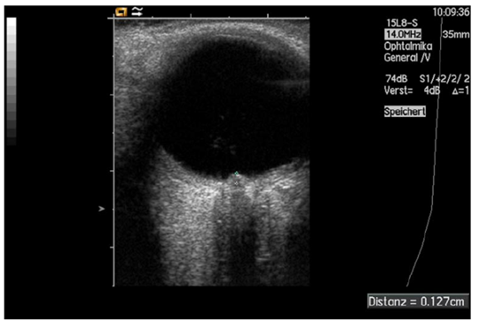
Fig. 8. Measurement of papilledema using ultrasound: disc elevation is quantified by putting the first caliper on the uppermost part of the swollen disc; the second caliper is positioned on the strongly reflecting line representing the lamina cribrosa 8