Archives for 2017

Neuro-ophthalmology Question of the Week: Bilateral Occipital Lobe Lesions & Visual Acuity

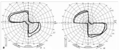
Question: When bilateral lesions of the retrochiasmal visual pathways produce a decrease in visual acuity which of the following are correct?
1. Such lesions produce symmetric VA loss in both eyes.
2. The VA loss from such lesions will be worse on the opposite side of the largest lesion.
3. The VA loss from such lesions will be worse on the same side of the largest lesion.
4. With the presence of retrochiasmal visual pathway lesions and different VA in the two eyes, an additional problem anterior to the chiasm must be present.
1

Neuro-ophthalmology Question of the Week: Optic Tract Defects

Question:
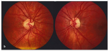
Which of the following are correct for a left optic tract lesion?
1. Right homonymous hemianopia
2. Left RAPD
3. Bowtie atrophy of the right optic nerve
4. Mostly temporal pallor of the left optic nerve

1

Neuro-ophthalmology Question of the Week: Ancillary Tests

Questions:
1. In the photostress test, how long does the patient look at a bright light held a few centimeters from the eye?
2. In the photostress test, what is the normal recovery time to within 1 line of best corrected visual acuity? 
3. What anatomic factor explains the RAPD with an optic tract lesion?
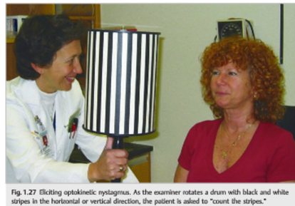
4. What are 5 clinical settings where OKN testing may be helpful?
5. Where is the lesion likely to be located in a patient with homonymous hemianopia and asymmetric OKN response?

1

Neuro-ophthalmology Question of the Week: Neuro-ophthalmology Coma Eye Exam

Question:
Which of the following are correct for a patient?
1. The eyes of a patient in coma will be closed.
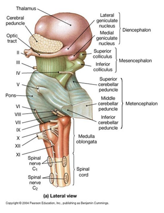
2. Because the pathways governing ocular motility traverse the entire brainstem, brainstem lesions will most often result in abnormal eye movements, and the lesion can be localized to the midbrain, pons, or medulla.
3. The presence of a dysconjugate ocular deviation in coma (horizontal, vertical, or oblique misalignment) often indicates a cranial nerve palsy or skew deviation.
4. Roving eye movements: slow ocular conjugate deviations in random directions indicate intact ocular motility function in the brainstem.
5. Periodic alternating (“ping-pong”) gaze: slow, repetitive, rhythmic, back-and-forth, horizontal conjugate eye movements indicates intact ocular motility function in the brainstem.
6. Normal vertical and horizontal oculocephalic reflexes indicate intact ocular motility function in the brainstem.

1

Neuro-ophthalmology Question of the Week: Optokinetic Testing

Question: In which of the following conditions is optokinetic stimulus testing useful?
1. 
Infantile nystagmus
2. Patients claiming complete blindness
3. Homonymous hemianopia
4. Internuclear ophthalmoplegia
5. Dorsal midbrain syndrome
6. Optic neuritis

1

Neuro-ophthalmology Question of the Week: Optic Neuropathy or Maculopathy

Question:
Which of the following clinical tests can help differentiate between optic neuropathy and maculopathy in a patient with a reduction in visual acuity?
1.Swinging flashlight pupillary reactions
2. Amsler Grid
3. Dilation lag in darkness
4. Anisocoria
5. Photostress recovery test
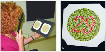
6. Color vision/saturation

1

https://neuro-ophthalmology.stanford.edu/2017/02/378/

Neuro-ophthalmology Question of the Week: Dilated pupil constricts to pilocarpine 0.125%

Question1:
A 35-year-old woman is evaluated for a 2-week history of photophobia and awareness that her right pupil is larger than the left. Examination shows absent right pupillary response to light stimulation of either eye. There is no diplopia and no evidence of extraocular muscle palsy. Upon local instillation of 0.125% pilocarpine, the larger pupil becomes smaller than the contralateral pupil.

Which of the following is the most likely location of the lesion?
1. ciliary ganglion
2. Edinger-Westphal nucleus
3. oculomotor nerve
4. optic chiasm
5. pretectal nucleus

2

Neuro-ophthalmology Question of the Week: Anisocoria – Horner syndrome & Heterochromia Iridis

Question: Which of the following are correct?
1. A heterochromia iridis-associated acquired Horner syndrome is usually due to a lesion of the postganglionic neuron (third order).
2. Heterochromia iridis-associated acquired Horner syndrome only occurs in children?
3. In a patient with isolated heterochromia iridis-associated Horner syndrome, the lighter eye is the one with the oculosympathetic paresis.
4. When both Waardenburg and congenital Horner syndromes occur in the same individual the darker iris is the one with the oculosympathetic paresis.

1

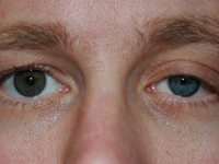
Neuro-ophthalmology Question of the Week: Horner Syndrome in Children

Question:
Which of the following are correct?
1. Heterochromia iridis in a child with Horner syndrome rules out neuroblastoma as a cause.
2. The most common cause of congenital Horner syndrome is a neuroblastoma.
3. Cervical neuroblastoma, a disease primarily of infants, has a favorable prognosis.
4. Cervical masses due to neuroblastoma, when felt, are commonly mistaken for infectious adenitis in children.
5. Cervical neuroblastoma in children almost always causes Horner syndrome.

1
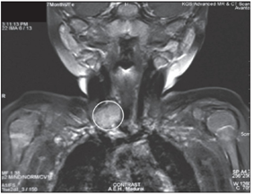
MRI showing a heterogeneously enhancing soft tissue lesion involving the root of neck

Neuro-ophthalmology Question of the Week: Anisocoria – Pseudo-Horner Syndrome

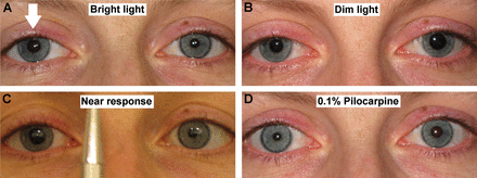
Question: Which of the following may reveal that the cause of anisocoria is not Horner syndrome?
1. Slit-lamp exam
2. The degree of anisocoria remains relatively constant in bright and dim illumination.
3. Repeat examinations of the patient on different days.
4. The absence of miosis on attempted near vision.
5. The failure of the pupil to constrict to light but does constrict with attempted near vision.

2