Teaching Video NeuroImages: Minimal anomalies of dorsal midbrain syndrome (Parinaud syndrome)

Teaching Video NeuroImages: Minimal anomalies of dorsal midbrain syndrome (Parinaud syndrome)
Pilar Rojas, Philippe Maeder and François-Xavier Borruat.
Neurology. January 03, 2017; 88 (1) RESIDENT AND FELLOW SECTION

Parinaud syndrome results from posterior commissure dysfunction, and is associated with 4 major signs: limitation of upgaze, pupillary light-near dissociation, convergence abnormalities, and Collier sign.1,2

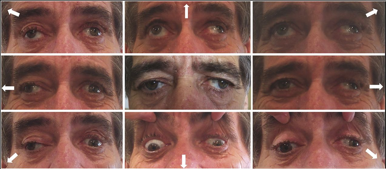
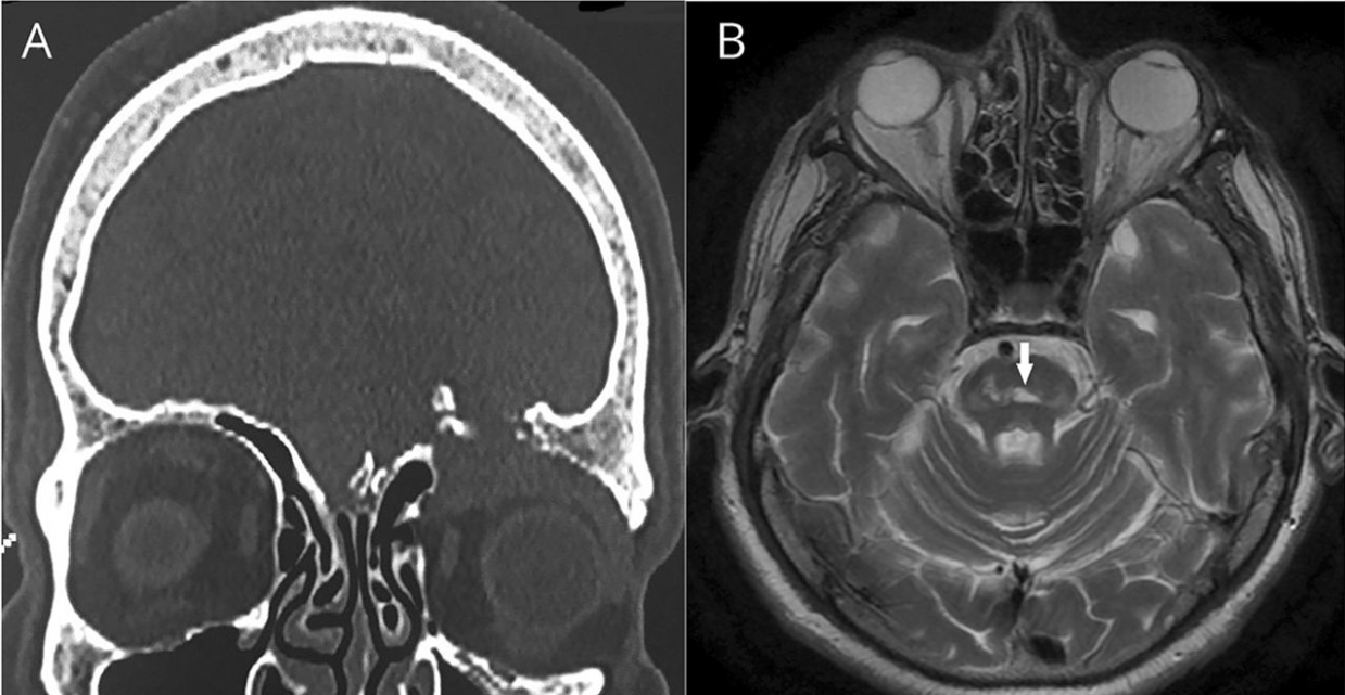
A 46-year-old man complained of vertical diplopia due to a subtle left skew deviation. Upgaze pursuit was normal, but upward saccades were slowed, without convergence abnormalities or Collier sign (video). Pupillary light-near dissociation was present (video). MRI revealed a tectal mesencephalic lesion (figure).
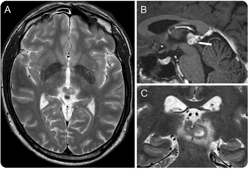
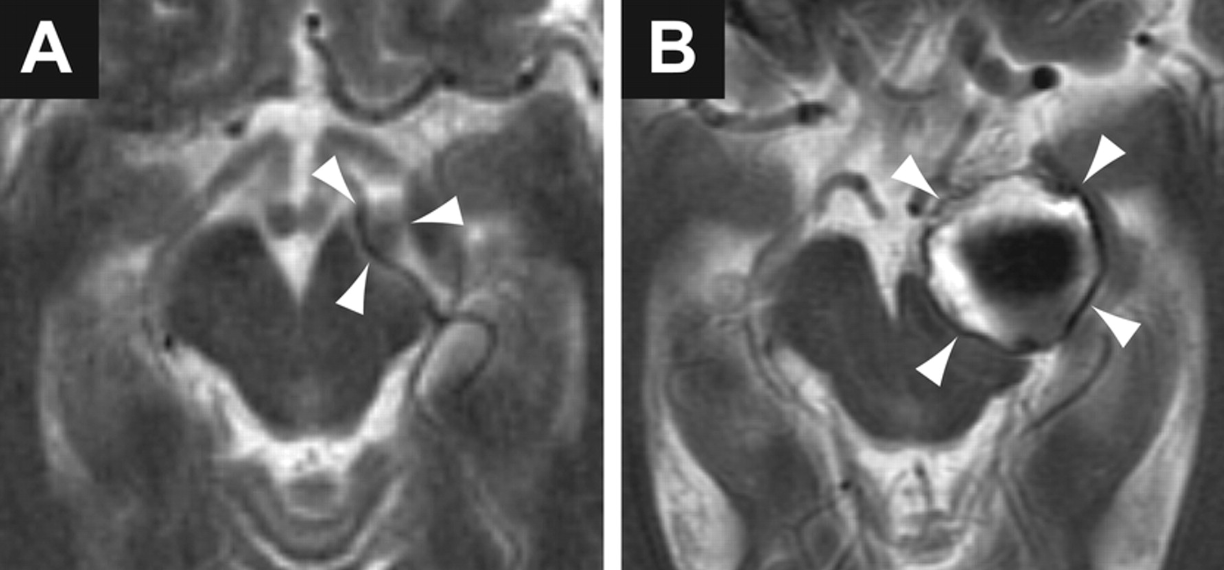
Figure Brain MRI
T2 axial and coronal cuts (A, C) and T1 sagittal cut with gadolinium (B) revealed a tumor originating from the walls of the third ventricle posteriorly, with an invasion of the posterior commissure (white arrow). The lesion was later biopsied and pathology revealed a pilocytic astrocytoma.

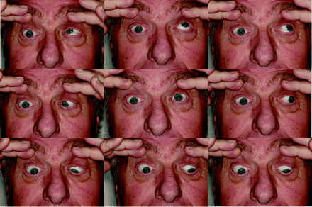
Video Ocular motility and pupils examination.
Horizontal pursuit and saccades, and both upward and downward vertical pursuit were normal in both eyes, namely without any limitation of upgaze. The speed of downward vertical saccades was normal, whereas upward saccades from primary gaze were slowed. The pupils were of intermediate and equal size, and pupillary light-near dissociation was present in both eyes.

Slowed upward saccades and pupillary light-near dissociation represent an early stage of posterior commissure dysfunction, before frank upgaze palsy, Collier sign, or convergence abnormalities.

AUTHOR CONTRIBUTIONS
Pilar Rojas is an author and contributed to drafting and revising the manuscript. Philippe Maeder is an author, contributed to data acquisition, and revised the manuscript. François-Xavier Borruat is an author, contributed to data acquisition, and revised the manuscript.

STUDY FUNDING No targeted funding reported.

DISCLOSURE The authors report no disclosures relevant to the manuscript. Go to Neurology.org for full disclosures.

Footnotes
  Supplemental data at Neurology.org
  Download teaching slides: Neurology.org

REFERENCES
1. Leigh RJ, Zee DS. The Neurology of Eye Movements, 3rd ed. part II, ch 10. Oxford: Oxford University Press; 1999:517–519.
2. Miller NR, Newman NJ. Clinical Neuro-Ophthalmology, 5th ed. vol 1, ch 29. Baltimore: Williams & Wilkins; 1998:1304–1311.


Teaching NeuroImage: Convergence spasm associated with midbrain compression by cerebral aneurysm

Teaching NeuroImage: Convergence spasm associated with midbrain compression by cerebral aneurysm
Konrad P. Weber, Matthew J. Thurtell, and G. Michael Halmagyi
Neurology. April 08, 2008; 70 (15) RESIDENT AND FELLOW SECTION

ARTICLE
A 72-year-old woman presented with intermittent diplopia. She had a history of vascular disease, but no history of stroke or psychiatric disturbance. On examination, she developed convergence spasm with associated miosis following fixation on near targets and during horizontal smooth pursuit (video, figure 1). The spasms were terminated with a blink. She had no ptosis, her eye movements were normal in range, and her pupils were equal in size with normal reactions to light. Neurologic examination was otherwise unremarkable. Five years prior to her current presentation, a left-sided terminal carotid artery aneurysm had been incidentally detected. The aneurysm gradually expanded to a diameter of 2.4 cm and compressed the midbrain (figure 2). The intermittent diplopia had developed in the months following endovascular coiling of the aneurysm.


Figure 1 (A–D) Convergence spasm precipitated by fixation on a near target and released after a blink (video, A).
(A) During fixation on a distant target, the patient has mid-range pupils.
(B) During fixation on a near target, the patient converges and has miotic pupils.
(C) After the near target is removed, convergence is maintained. The pupils remain miotic, which helps to differentiate from bilateral sixth nerve palsies.
(D) After a blink, the patient is able to re-fixate on the distant target and the pupils return to mid-range diameter.
(E) Binocular search coil recording during sinusoidal horizontal smooth pursuit, in which there is a period of convergence spasm that is released by a blink (video, B).
(F) The vergence angle progressively increases up to 50° during the period of convergence spasm (gray triangle).


Figure 2 T2-weighted axial MRI demonstrating progressive compression of the midbrain by an expanding aneurysm (arrowheads) arising from the terminal left carotid artery
(A) MRI 5 years prior to onset of symptoms.
(B) MRI at the time of presentation with convergence spasm (after endovascular coiling of the aneurysm).

Convergence spasm is characterized by the inappropriate appearance of the near triad, which consists of convergence, miosis, and accommodation.1 Most patients are young and the cause is psychiatric; a focal lesion is rarely found.1 Since the neural substrate for the near triad is located in the midbrain, convergence spasm could occur with a midbrain lesion. In our patient, isolated convergence spasm may have been a manifestation of midbrain compression. Thus, in patients presenting with convergence spasm, the presence of other focal neurologic signs or the absence of psychiatric history, especially in the elderly, should prompt neuroimaging. Furthermore, it is important not to mistake convergence spasm for bilateral sixth nerve palsies; miosis on attempted lateral gaze is diagnostic of convergence spasm.2

ACKNOWLEDGMENT The authors thank Dr. Geoffrey D. Parker for help with the interpretation of the MRI.

Footnotes
   Supplemental data at www.neurology.org
   
Supported by Garnett Passe and Rodney Williams Memorial Foundation.
   Disclosure: The authors report no conflicts of interest.
   Series editor: Mitchell S.V. Elkind MD, MS, Section Editor

REFERENCES
1. Goldstein JH, Schneekloth BB. Spasm of the near reflex: a spectrum of anomalies. Surv Ophthalmol. 1996;40:269–278
2. Griffin JF, Wray SH, Anderson DP. Misdiagnosis of spasm of the near reflex. Neurology. 1976;26:1018–1020


Teaching Video NeuroImages: Pulsatile proptosis and wall-eyed bilateral internuclear ophthalmoplegia

Teaching Video NeuroImages: Pulsatile proptosis and wall-eyed bilateral internuclear ophthalmoplegia
Eleni Papageorgiou, Evangelia E. Tsironi, Sofia Androudi and Petros Koltsidopoulos
Neurology. April 24, 2018; 90 (17) RESIDENT & FELLOW SECTION

ARTICLE
A 65-year-old man with a history of hypertension presented with double vision. Examination revealed wall-eyed bilateral internuclear ophthalmoplegia (WEBINO) (figure 1) and nonsymptomatic, pulse-synchronous, pulsatile proptosis of the left eye (video, links.lww.com/WNL/A389). According to the patient, there was a history of blunt head trauma in childhood and the pulsatile eye was present since then. CT revealed a meningocele into the left orbit due to a bony defect in the orbital roof (figure 2A). CT angiography ruled out a carotid-cavernous fistula. MRI showed an acute infarction at the midline of the midbrain tegmentum, which involved the medial longitudinal fasciculus bilaterally and caused the WEBINO (figure 2B).1 The pulsatile proptosis was attributed to the childhood head injury and due to the orbital roof fracture and associated meningocele pulsation of the brain vessels passed onto the CSF.2

Figure 1 Extraocular movements in 9 cardinal gaze positions
Extraocular eye movements demonstrate exotropia of both eyes in primary positions and bilateral internuclear ophthalmoplegia with impaired adduction on attempted lateral gaze. Upgaze is also mildly affected.

Figure 2 Coronal brain CT and axial brain MRI
(A) Coronal brain CT shows fracture of the left orbital roof with an associated meningocele.
(B) T2-weighted axial brain MRI demonstrates an acute infarction at the midline of the midbrain tegmentum, which involves the medial longitudinal fasciculus bilaterally.

Author contributions E.P.: study concept and design, acquisition of data, analysis and interpretation, writing of the manuscript. E.E.T. and S.A.: study supervision, critical revision of the manuscript for important intellectual content. P.K.: acquisition of data, analysis and interpretation.

Study funding No targeted funding reported.

Disclosure The authors report no disclosures relevant to the manuscript.

References:
1. Kim JS, Jeong SH, Oh YM, Yang YS, Kim SY. Teaching NeuroImage: wall-eyed bilateral internuclear ophthalmoplegia (WEBINO) from midbrain infarction. Neurology 2008;70:e35.
2. Chousterman B, Gualino V, Dohan A, Payen D. A mysterious post-traumatic pulsatile exophthalmos. Intens Care Med 2014;40:102–103.


Clinical Reasoning: A 64-year-old man with painful, unilateral external ophthalmoplegia

Clinical Reasoning: A 64-year-old man with painful, unilateral external ophthalmoplegia
M. Tariq Bhatti. Neurology. 2010; 75 (8)
RESIDENT AND FELLOW SECTION

SECTION 1
A 64-year-old man was referred for evaluation of double vision in March 2009. Four months prior, he experienced daily pain in the region of the right forehead and right eye. Two months later, he noticed constant binocular, vertical double vision. Evaluation by an outside ophthalmologist resulted in the diagnosis of a right 4th cranial nerve (CN) palsy, a normal cranial and orbital MRI study with contrast, and an unremarkable laboratory evaluation. One month later, his right eye began “turning in,” and within a few days he was unable to abduct the eye. Several weeks later, the right eyelid began to droop and progressively worsened over the next several days to complete closure of the eye.

His past medical history was notable for arterial hypertension, depression, rheumatoid arthritis, gastroesophageal reflux disease, nephrolithiasis, squamous cell carcinoma of the forehead, and a precancerous melanoma of the left ear.

When he was seen in the neuro-ophthalmology clinic, visual acuity was 20/20 in each eye. Color vision was intact in each eye. The right pupil was 2 mm larger than the left and was nonreactive to light or near effort. There was no relative afferent pupillary defect (RAPD). Dilated fundus examination was normal in each eye. Eye movements of the right eye were limited in all directions and there was complete right upper eyelid ptosis (figure 1). Corneal sensation of the right eye was absent and there was numbness over the right forehead. The remainder of the cranial nerve examination was normal.

Figure1

Figure 1 Nine cardinal positions of eye movements
There is limited movement of the right eye in all directions of gaze. There was no intorsion of the right eye on attempted downgaze. The right eyelid is manually elevated because of the complete right eyelid ptosis (center, middle panel). Permission obtained from patient.

Questions for consideration:
1. What is the clinical presentation?
2. Where does the lesion localize?

Teaching NeuroImages: Upright-supine test to evaluate vertical diplopia

Teaching NeuroImages: Upright-supine test to evaluate vertical diplopia
Nailyn Rasool and Sashank Prasad
Neurology May 12, 2015; 84 (19)
RESIDENT AND FELLOW SECTION

Article:
A 36-year-old woman presented with vertical diplopia, nausea, and disequilibrium. Maddox rod testing was performed in the upright and supine positions (figures 1 and 2).


Figure 1 
Upright-supine test
During Maddox rod testing, the patient used her fingers to demonstrate the separation of images.
The vertical deviation decreased substantially (over 50%) when supine compared to upright.

Teaching Video NeuroImages: Bilateral abducens ocular neuromyotonia

Teaching Video NeuroImages: Bilateral abducens ocular neuromyotonia
Kavin Vanikieti and Joseph F. Rizzo
Neurology September 05, 2017; 89 (10)
RESIDENT AND FELLOW SECTION http://n.neurology.org/content/89/10/e128

A 44-year-old woman with a history of resolved nasopharyngeal carcinoma presented with an 8-month history of transient binocular horizontal diplopia. She received complete concurrent chemoradiotherapy 7 years ago. Following prolonged eccentric gaze to the right, she developed involuntary contraction of right lateral rectus, which resulted in 60 seconds of right exotropia. These events also occurred following prolonged eccentric gaze to the left, which resulted in left exotropia (video 1). Treatment with carbamazepine improved her symptoms (video 2). Ocular neuromyotonia should be included as a differential diagnosis of transient diplopia, especially in those who have undergone prior radiotherapy to the parasellar region.1

Video 1 – Pretreatment
Following prolonged eccentric gaze to the right, the patient developed involuntary contraction of right lateral rectus, which resulted in right exotropia. Each episode lasted approximately 60 seconds and then the eyes returned to normal alignment. These events also occurred following prolonged eccentric gaze to the left, which resulted in left exotropia.

Video 2 – Post Treatment
Complete resolution with carbamazepine

Teaching SlidesPowerPoint Presentation

Reference: 1. Leigh RJ, Zee DS. The Neurology of Eye Movements, 5th ed. Oxford: Oxford University Press; 2015:615.

Clinical Reasoning: A 22-year-old man with diplopia

Clinical Reasoning: A 22-year-old man with diplopia
Chelsea Meyer, DO, Don Raphael P. Wynn, MD, Stefan M. Pulst, MD, DrMed Ricky Chen, MD, Kathleen Digre, MD Correspondence to Dr. Meyer: chelsea.meyer@hsc.utah.edu
Neurology October 11, 2016; 87 (15)
RESIDENT & FELLOW SECTION Section Editor John J. Millichap, MD

SECTION 1
A 22-year-old previously healthy man presented to an ophthalmology clinic with binocular horizontal diplopia. He had recently traveled to the main island of Hawaii. About 2 weeks after returning home, he developed a severe headache with associated fever, emesis, photophobia, phonophobia, and neck stiffness. He also reported a sensation of pressure in his left eye and both ears but denied any pulsatile tinnitus or transient vision loss. Over the next 2 weeks, his headaches worsened, causing him to wake up frequently in the night. He then developed horizontal diplopia that was worse at a distance and was referred to the neuro-ophthalmology clinic. Examination. The patient had some limitation in neck flexion with associated pain. His visual acuity was 20/20 on the right and 20/25 on the left. Pupillary examination showed no relative afferent defect and visual fields were full. He was found to have bilateral abducens palsy with an esotropia of 30 D and a left hypertropia of 2 D with notable mild right head tilt. The hypertropia was thought to be a partial left 4th nerve palsy with a compensatory right head tilt, although a full Parks-Bielschowsky 3-step test was not preformed to confirm this. He had moderate to severe papilledema on funduscopic examination (figure, A). The remainder of his neurologic and ophthalmologic examination was within normal limits.


Figure (A) Funduscopic photographs of the optic disc of the left eye show papilledema with blurring of the disc margin, optic nerve hyperemia, and peripapillary hemorrhages.

Questions for consideration:
1. Given his papilledema and bilateral abducens nerve palsy, where would you localize this?
2. What is the differential diagnosis for bilateral abducens nerve palsy?

Clinical Reasoning: A 20-year-old man with headache and double vision

Khaled Moussawi, Anoopum Gupta and Haatem Reda
Neurology October 11, 2016; 87 (15)
Correspondence to Dr. Moussawi: khaled.moussawi@nih.gov
RESIDENT AND FELLOW SECTION
Section Editor John J. Millichap, MD

From Harvard Medical School (K.M., A.G., H.R.), Boston; Department of Neurology (K.M., A.G., H.R.), Massachusetts General Hospital, Boston; and Department of Neurology (K.M., A.G.), Brigham and Women’s Hospital, Boston, MA. Dr. Moussawi is currently with the National Institute on Drug Abuse; and the Department of Psychiatry, Johns Hopkins Medicine, Baltimore, MD.

SECTION 1
A 20-year-old man presented to the emergency department with 1 week of headaches and double vision following 2 days of fever (1028 F), nausea, and vomiting. His headache was progressively worsening, throbbing behind his right eye, nonpositional, and associated with photophobia, blurry vision, and pain with eye movement. Occasionally, it was severe enough to wake him up from sleep. Horizontal double vision ensued soon after the headache. His diplopia was worse looking at a distance, improved on leftward gaze, worsened on rightward gaze, and resolved with closing one eye. He denied neck stiffness, focal weakness, numbness, or other neurologic symptoms. He denied recent rashes, infections, or tick bites. He lives on a farm in central Brazil. He arrived in Massachusetts 2 months before his presentation to visit family members. He had no significant medical history and took no medications. He did not smoke or use drugs.

Question for consideration:
1. What is the localization of his presenting symptom?

Clinical Reasoning: Two see or not two see—Is it really double vision?

Clinical Reasoning: Two see or not two see—Is it really double vision?
Richard Ronan Murphy, MBChB Abdullah Al Sawaf, MD Danny R. Rose Jr., MD Larry B. Goldstein, MD Charles D. Smith, MD
Neurology  August 08, 2017; 89 (6)
RESIDENT AND FELLOW SECTION
RESIDENT & FELLOW SECTION
Section Editor John J. Millichap, MD
Correspondence to R.R. Murphy: ronan.murphy@uky.edu

SECTION 1
A 57-year-old right-handed woman presented to the emergency department with complaints of double vision and intractable nausea that began abruptly 2 days earlier. Her visual symptoms were characterized as seeing overlapping or separate horizontally or diagonally displaced objects. She had no history of headaches or stroke. Her cerebrovascular risk factors included hypertension, type II diabetes, coronary artery disease, and cigarette smoking. Her medications included clopidogrel, lisinopril, paroxetine, and oxycodone. Her family history was notable for late-onset ischemic heart disease in her parents with no first-degree relatives with early vascular disease. On examination, her blood pressure was 158/101 mm Hg, pulse rate was 87 bpm, and she was afebrile. She was alert and fully oriented. Her attention, recall of recent events, and general fund of knowledge were normal. Her speech was fluent and nondysarthric. Cranial nerve examination was notable for no dysconjugacy or nystagmus, but double vision predominately in the horizontal plane, in all directions of gaze. The diplopia persisted with monocular vision in each eye, and did not improve with a pinhole test. The degree of diplopia waxed and waned during the examination, with visual field extinction tests being difficult to perform reliably. Her pupils were equal with bilateral hippus. Visual fields were full to confrontation. Direct funduscopy revealed normal optic discs. She had a mild right hemiparesis with mild right arm and leg drift, but no facial asymmetry. There was mild hypesthesia over her right arm and leg and appendicular ataxia in her right arm that was worse with eyes open. She did not have extinction to double simultaneous sensory stimuli. Gait evaluation was deferred during her initial examination.

Questions for consideration:
1. What is the significance of the presence of diplopia in both eyes, with either eye closed?
2. What more could be elicited from the history and examination to help characterize the problem?

Heimann-Bielschowsky phenomenon

Recommended Reading
Teaching Video NeuroImages: Heimann-Bielschowsky phenomenon
A harmless monocular nystagmus

Article:
Heimann-Bielschowsky phenomenon (HBP) is a rare form of dissociated nystagmus.1 A 38-year-old man complained of poor vision in his right eye for 6 years. Right visual acuity was 20/200 due to traumatic aphakia (cataract extraction without intraocular lens) and 20/20 in the left eye. An asymptomatic coarse, low frequency, pendular, vertical nystagmus was manifest in the right eye only (video, http://links.lww.com/WNL/A177). HBP is an asymptomatic monocular slow, pendular, mostly vertical nystagmus, which can develop years after uniocular severe visual loss.1 The pathogenesis of HBP is debated, but vertical fusion disruption due to monocular visual loss is hypothesized.2 Recognizing HBP should prevent unnecessary investigations or treatments.

Teaching slides: http://links.lww.com/WNL/A178

Video: http://links.lww.com/WNL/A177

Neurology
February 20, 2018; 90 (8)
RESIDENT & FELLOW SECTION

References

  1. Yee RD, Jelks GW, Baloh RW, Honrubia V. Uniocular nystagmus in monocular visual loss. Ophthalmology 1979;86:511–522.
  2. Leigh RJ, Thurston SE, Tomsak RL, Grossman GE, Lanska DJ. Effect of monocular visual loss upon stability of gaze. Invest Ophthalmol Vis Sci 1989;30:288–292.