Recommended Reading – The expanding burden of idiopathic intracranial hypertension

Recommended Reading – The expanding burden of idiopathic intracranial hypertension

The expanding burden of idiopathic intracranial hypertension
Susan P. Mollan, Magda Aguiar, Felicity Evison, Emma Frew & Alexandra J. Sinclair. Eye (2018)

Free Full Text: https://www.nature.com/articles/s41433-018-0238-5

Abstract
OBJECTIVE: To quantify the hospital burden and health economic impact of idiopathic intracranial hypertension.

METHODS: Hospital Episode Statistics (HES) national data was extracted between 1st January 2002 and 31st December 2016. All those within England with a diagnosis of idiopathic intracranial hypertension were included. Those with secondary causes of raised intracranial pressure such as tumours, hydrocephalus and cerebral venous sinus thrombosis were excluded.

RESULTS: A total of 23,182 new IIH cases were diagnosed. Fifty-two percent resided in the most socially deprived areas (quintiles 1 and 2). Incidence rose between 2002 and 2016 from 2.3 to 4.7 per 100,000 in the general population. Peak incidence occurred in females aged 25 (15.2 per 100,000). 91.6% were treated medically, 7.6% had a cerebrospinal fluid diversion procedure, 0.7% underwent bariatric surgery and 0.1% had optic nerve sheath fenestration. Elective caesarean sections rates were significantly higher in IIH (16%) compared to the general population (9%), p < 0.005. Admission rates rose by 442% between 2002 and 2014, with 38% having repeated admissions in the year following diagnosis. Duration of hospital admission was 2.7 days (8.8 days for those having CSF diversion procedures). Costs rose from £9.2 to £50 million per annum over the study period with costs forecasts of £462 million per annum by 2030.

CONCLUSIONS: IIH incidence is rising (by greater than 100% over the study), highest in areas of social deprivation and mirroring obesity trends. Readmissions rates are high and growing yearly. The escalating population and financial burden of IIH has wide reaching implications for the health care system.

Composite figure.


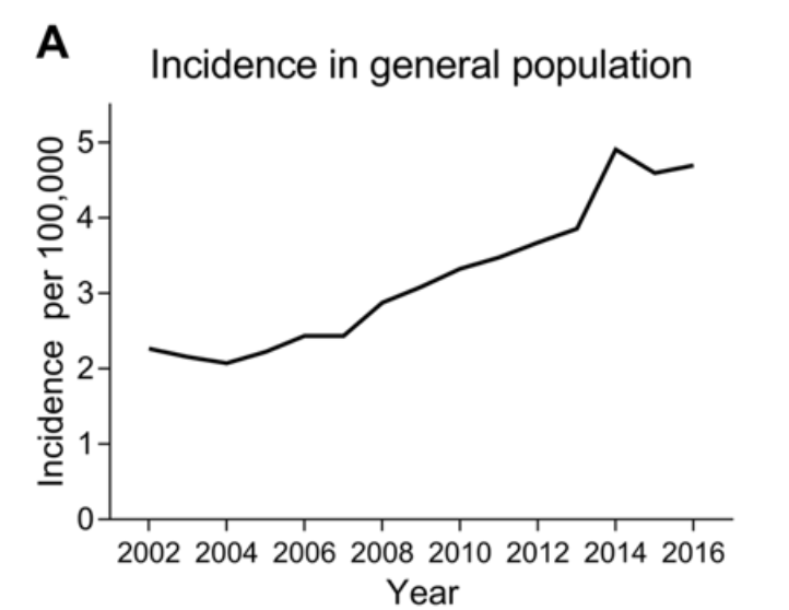
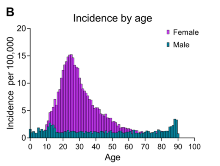
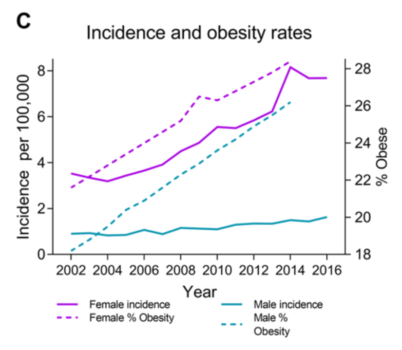
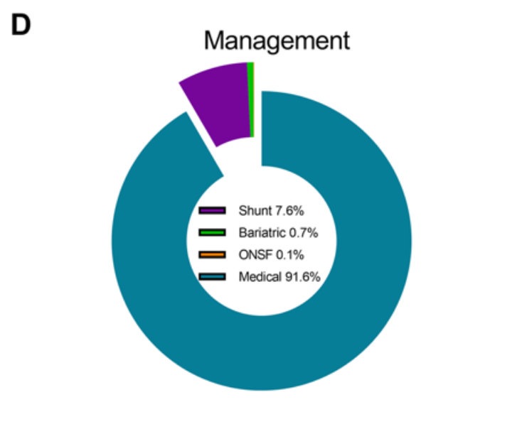
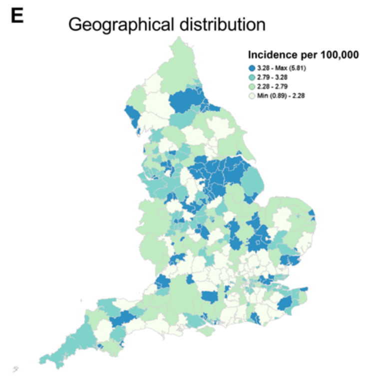
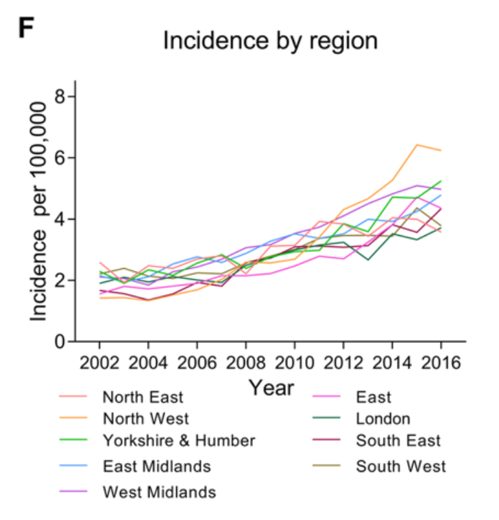
a Incidence in the general population. b Incidence by age and gender. c Annual incidence in females and males and Obesity rates (% obesity per annum (body mass index ≥ 30), age-standardized in 18 years + by gender in the United Kingdom. From World Health organisation http://apps.who.int/gho/data/node.main.A900A?lang=en Accessed 6 Oct 2017. d Management of IIH in the cohort. e Geographical distribution of diagnosed cases of IIH in England. F Distribution of cases by region per annum.


Recommended Reading – Toxic medications in Leber’s hereditary optic neuropathy

Recommended Reading – Toxic medications in Leber’s hereditary optic neuropathy

Toxic medications in Leber’s hereditary optic neuropathy.
Kogachi K, Ter-Zakarian A, Asanad S, Sadun A, Karanjia R
Mitochondrion. 2018 Aug 4. pii: S1567-7249(18)30036-9. doi: 10.1016/j.mito.2018.07.007. [Epub ahead of print]

Abstract
Leber’s hereditary optic neuropathy (LHON) is a maternally inherited mitochondrial disorder characterized by acute bilateral vision loss. The pathophysiology involves reactive oxygen species (ROS), which can be affected by medications. This article reviews the evidence for medications with demonstrated and theoretical effects on mitochondrial function, specifically in relation to increased ROS production. The data reviewed provides guidance when selecting medications for individuals with LHON mutations (carriers) and are susceptible to conversion to affected. However, as with all medications, the proven benefits of these therapies must be weighed against, in some cases, purely theoretical risks for this unique patient population.

_______________________________

… In this article, we review medications that are proposed to increase ROS. Given the association between ROS and conversion, understanding the effects of these agents on mitochondria may assist clinicians to understand the implications of these classes of medications for patients with LHON. Medications on this list can be used in patients with LHON, especially when no alternative exists. As with all therapies, a balance must be struck between the therapeutic benefits and potential harm, which may warrant special consideration for patients with LHON.

Full Text: https://drive.google.com/open?id=12HvHSKJaRj7WOvkJQKWlj8FOVbcSoNbj

Recommended Reading – Abnormal Eye Movements in Paraneoplastic Syndromes

Recommended Reading – Abnormal Eye Movements in Paraneoplastic Syndromes

Opsoclonus (Ri antibodies)
931-1 Paraneoplastic Opsoclonus http://www.kaltura.com/index.php/extwidget/preview/partner_id/797802/uiconf_id/27472092/entry_id/0_47rqop2q/embed/auto?
Title: Paraneoplastic Opsoclonus Subject: Opsoclonus; Ocular Flutter; Oscillopsia; Saccadic Oscillations; Paraneoplastic Cerebellar Syndrome; Adenocarcinoma of the Breast; Anti-Ri Antibody; Paraneoplastic Opsoclonus; Paraneoplastic Ocular Flutter; Saccadomania Description: This patient is the index case of the Anti-Ri antibody, published in Annals of Neurology in 1988 (4). The Anti-Ri antibody is recognized to be a paraneoplastic marker in patients with breast and gynecological malignancies (10). The history of this case is particularly important because she was initially misdiagnosed as a case of acute labyrinthitis.

Ocular flutter (Ri antibodies)
https://www.aao.org/annual-meeting-video/ocular-flutter
Rod Foroozan. AAO Subspecialty Day 2011: Neuro-Ophthalmology (AAO sign-in may be required)
In this case discussion from Neuro-Ophthalmology Subspecialty Day 2011, Dr. Rod Foroozan presents a case of ocular flutter, a rare disorder characterized by back-to-back saccades. The abnormal ocular oscillations are believed to result from dysfunction of the pause/burst cells, which govern saccade generation, in the pons. The most common causes can be categorized as inflammatory, infectious or paraneoplastic. Most patients with an infectious cause show spontaneous improvement weeks to months after the onset of symptoms.

Cerebellar degeneration (Yo antibodies)
Paraneoplastic Cerebellar Degeneration. Hain TC. 2016
Downbeat Nystagmus anti-YO paraneoplastic cerebellar degeneration
Downbeat Nystagmus in paraneoplastic cerebellar degeneration in left gaze
Downbeat Nystagmus in paraneoplastic cerebellar degeneration increased with convergence
Website https://www.dizziness-and-balance.com/disorders/central/cerebellar/pcd.htm

Downbeat Nystagmus  https://www.youtube.com/watch?v=FrlsK4MF3Ao
Downbeat Nystagmus is a “central” or brain-related abnormal eye movement that could have no identifiable cause, but it might suggest abnormalities in the brainstem or cerebellum region of the central nervous system, is often seen in cerebellar degeneration syndromes, vitamin B12 or B1 (thiamine) deficiency, magnesium deficiency,  and may be part of a paraneoplastic syndrome.

Elliptical Nystagmus and Oscillopsia
Teaching Video NeuroImages: P/Q-type voltage-gated calcium channel–associated paraneoplastic elliptical nystagmus. Mistry EA, Lee AG, Lai EC. Neurology. 2016.
A 71-year-old chronic smoker had an 11-month history of monocular followed by binocular elliptical nystagmus and oscillopsia (video at Neurology.org). MRI brain showed extensive periventricular T2 signal changes (figure) and CSF showed elevated protein to 102 mg/dL. CSF and serum paraneoplastic panel revealed elevated serum titers of anti-P/Q-type voltage-gated calcium channel (VGCC) and anti-neuronal-type voltage-gated potassium channel antibodies. An underlying malignancy was not found after an extensive investigation. The patient was treated with carbamazepine for symptomatic control, followed by high-dose IV methylprednisolone, resulting in moderate improvement. Anti-VGCC antibodies have been implicated in paraneoplastic nystagmus and small cell lung cancer is the most common associated malignancy.1,2

Paraneoplastic upbeat nystagmus.

Wray SH, Martinez-Hernandez E, Dalmau J, Maheshwari A, Chen A, King S, Bishop Pitman M, Leigh RJ. Neurology.2011:16;77(7):691-3.
…During attempted fixation of a far target, she had prominent UBN, lid nystagmus, and saccadic intrusions (video on the Neurology® Web site at www.neurology.org). UBN suppressed during near viewing and showed marked dependency on head position, upbeat when erect, absent when supine, reduced when prone, and beating away from the ground (apogeotropic) when lying on either side.
A CT-guided core biopsy revealed a pancreatic endocrine neoplasm.
Paraneoplastic antibody testing, including anti-Ri, Anti-Yo, anti-Hu, anti-Ma1 and Ma2, anti-ZiC4, and anti-CV2, was positive for anti-Hu antibodies at a titer of 1/15,360. The tumor showed robust reactivity with a monoclonal antibody against Hu confirming the expression of this antigen (figure, A and B). Analysis of patient’s serum and CSF for antibodies against the neuropil of brain, brainstem, and cerebellum (usually indicating a cell membrane or cell surface autoantigen) showed both samples had reactivity with the nuclei of neurons (Hu antigen) as well as with an unknown antigen expressed in the neuropil of brain.

Figure

Expression of Hu antigen by the patient’s tumor, and demonstration of an antibody against a neuronal cell surface antigen
The tumor shows intense reactivity with a mouse monoclonal antibody against human Hu (Molecular Probes; Eugene, OR; Cat# A-21271) used at dilution 1:50 (A).
As a contrast, the tumor does not show reactivity with normal mouse immunoglobulin G used at the same dilution (control section, B).
The immunostaining was performed using the avidin-biotin-peroxidase method followed by hematoxylin counterstaining (×200). Using cultures of dissociated rat hippocampal neurons patient’s serum (diluted 1:100) shows reactivity with the neuronal cell surface (C). The immunolabeling was done using live, not permeabilized neurons, as reported.7
Full-Text https://www.ncbi.nlm.nih.gov/pmc/articles/PMC3159092/

Paraneoplastic disorders may also cause slow saccades and limited vertical movements (Hu, Ma/Ta antibodies).

A recent review:  Paraneoplastic Neurologic Syndromes. Rosenfeld MR &  Josep Dalmau J. Neurol Clin 36 (2018) 675–685

Recommended Reading – Vitamin D for the management of multiple sclerosis.

Recommended Reading – Vitamin D for the management of multiple sclerosis.
Jagannath VA1, Filippini G, Di Pietrantonj C, Asokan GV, Robak EW, Whamond L, Robinson SA.
Cochrane Database Syst Rev. 2018 Sep 24;9:CD008422.
doi: 10.1002/14651858.CD008422.pub3.

Abstract
BACKGROUND:
This review is an update of a previously published review, “Vitamin D for the management of multiple sclerosis” (published in the Cochrane Library; 2010, Issue 12). Multiple sclerosis (MS) is characterised by inflammation, demyelination, axonal or neuronal loss, and astrocytic gliosis in the central nervous system (CNS), which can result in varying levels of disability. Some studies have provided evidence showing an association of MS with low levels of vitamin D and benefit derived from its supplementation.

OBJECTIVES:
To evaluate the benefit and safety of vitamin D supplementation for reducing disease activity in people with MS.

SEARCH METHODS:
We searched the Cochrane Multiple Sclerosis and Rare Diseases of the CNS Specialized Register up to 2 October 2017 through contact with the Information Specialist with search terms relevant to this review. We included references identified from comprehensive electronic database searches and from hand searches of relevant journals and abstract books from conferences.

SELECTION CRITERIA:
We included randomised controlled trials (RCTs) and quasi-RCTs that compared vitamin D versus placebo, routine care, or low doses of vitamin D in patients with MS. Vitamin D was administered as monotherapy or in combination with calcium. Concomitant interventions were allowed if they were used equally in all trial intervention groups.

DATA COLLECTION AND ANALYSIS:
Two review authors independently extracted data and assessed the methodological quality of studies, while another review author sorted any disagreements. We expressed treatment effects as mean differences (MDs) for continuous outcomes (Expanded Disability Status Scale and number of magnetic resonance imaging (MRI) gadolinium-enhancing T1 lesions), as standardised MDs for health-related quality of life, as rate differences for annualised relapse rates, and as risk differences (RDs) for serious adverse events and minor adverse events, together with 95% confidence intervals (CIs).

MAIN RESULTS:
We identified 12 RCTs enrolling 933 participants with MS; 464 were randomised to the vitamin D group, and 469 to the comparator group. Eleven trials tested vitamin D₃, and one trial tested vitamin D₂. Vitamin D₃ had no effect on the annualised relapse rate at 52 weeks’ follow-up (rate difference -0.05, 95% CI -0.17 to 0.07; I² = 38%; five trials; 417 participants; very low-quality evidence according to the GRADE instrument); on the Expanded Disability Status Scale at 52 weeks’ follow-up (MD -0.25, 95% CI -0.61 to 0.10; I² = 35%; five trials; 221 participants; very low-quality evidence according to GRADE); and on MRI gadolinium-enhancing T1 lesions at 52 weeks’ follow-up (MD 0.02, 95% CI -0.45 to 0.48; I² = 12%; two trials; 256 participants; very low-quality evidence according to GRADE). Vitamin D₃ did not increase the risk of serious adverse effects within a range of 26 to 52 weeks’ follow-up (RD 0.01, 95% CI -0.03 to 0.04; I² = 35%; eight trials; 621 participants; low-quality evidence according to GRADE) or minor adverse effects within a range of 26 to 96 weeks’ follow-up (RD 0.02, 95% CI -0.02 to 0.06; I² = 20%; eight trials; 701 participants; low-quality evidence according to GRADE). Three studies reported health-related quality of life (HRQOL) using different HRQOL scales. One study reported that vitamin D improved ratings on the psychological and social components of the HRQOL scale but had no effects on the physical components. The other two studies found no effect of vitamin D on HRQOL. Two studies reported fatigue using different scales. One study (158 participants) reported that vitamin D₃ reduced fatigue compared with placebo at 26 weeks’ follow-up. The other study (71 participants) found no effect on fatigue at 96 weeks’ follow-up. Seven studies reported on cytokine levels, four on T-lymphocyte proliferation, and one on matrix metalloproteinase levels, with no consistent pattern of change in these immunological outcomes. The randomised trials included in this review provided no data on time to first treated relapse, number of participants requiring hospitalisation owing to progression of the disease, proportion of participants who remained relapse-free, cognitive function, or psychological symptoms.

AUTHORS’ CONCLUSIONS:
To date, very low-quality evidence suggests no benefit of vitamin D for patient-important outcomes among people with MS. Vitamin D appears to have no effect on recurrence of relapse, worsening of disability measured by the Expanded Disability Status Scale (EDSS), and MRI lesions. Effects on health-related quality of life and fatigue are unclear. Vitamin D₃ at the doses and treatment durations used in the included trials appears to be safe, although available data are limited. Seven ongoing studies will likely provide further evidence that can be included in a future update of this review.

Update of Vitamin D for the management of multiple sclerosis. [Cochrane Database Syst Rev. 2010]

Full Text https://drive.google.com/open?id=1_ESVaPi-Rea_BzZ7E-EbJp-y7GR_6ZMU

Recommended Reading – Giant Cell Arteritis Treatment – Anti-Interleukin-6 Antibody

Recommended Reading – Giant Cell Arteritis Treatment – Anti-Interleukin-6 Antibody

IL-6 Blockade and its Therapeutic Success in Giant Cell Arteritis
Sebastian Unizony, MD, Tanaz A. Kermani, MD, MS: J Neuro-Ophthalmol 2018; 00: 1-8
CONCLUSIONS AND FUTURE PERSPECTIVES
Until recently, therapies that could maintain disease remission and prevent the well-known toxicity associated with excessive CS exposures have been the greatest unmet need for the GCA population. Studies elucidating the role of IL-6 in the inflammatory cascade in general and in the pathogenesis of GCA in particular have been instrumental in the eventual success of IL-6 blockade for the treatment of this condition. Further research is now required to answer several outstanding questions pertaining to the duration of TCZ treatment, the use of CS in patients receiving TCZ (e.g., can TCZ be used in monotherapy?), and whether TCZ is effective in controlling arterial inflammation and preventing large-artery complications. These and other questions will fine tune the use of TCZ for GCA. In addition, IL-6 inhibition has made even more pressing the need for accurate biomarkers to monitor disease activity and response to treatment.

More than 25 years passed from the initial observations that patients with GCA demonstrate an increased IL-6 signal to the demonstration of the efficacy of IL-6 blockade in rigorous clinical trials. We should do better. Fortunately, our understanding of the mechanisms of disease involved in GCA has improved and will likely continue to evolve in the future leading to the discovery of other important pathways and targeted treatment strategies

Full Text https://drive.google.com/open?id=1RUM9u7PRCekCpiDj-l9Ksf2eLCqne3xU  


Anti-Interleukin-6 Antibody as Treatment for Giant Cell Arteritis
Joyce Liao: J Neuro-Ophthalmol 2018; 00: 1-3
Choosing the right treatment for patients with GCA remains challenging. Although there is now an approved alternative to corticosteroids, our experience with TCZ is still limited compared with more than 6 decades of experience with corticosteroids. TCZ has promising efficacy in GCA, but there have been several reports of clinically significant, albeit rare, side effects such as severe neutropenia, recurrent pneumonia, cytomegalovirus, and other infections (30–32). Further research into the pathogenesis of GCA and the development of new therapies that can reverse vision loss and prevent relapse are critically needed.

Full Text https://drive.google.com/open?id=1gW9gf4HKGtg6onQtKliREMb2mYR9otzc


Recommended Reading – Terson Syndrome

Recommended Reading – Terson Syndrome
Terson syndrome in subarachnoid hemorrhage, intracerebral hemorrhage, and traumatic brain injury. Czorlich P, Skevas C, Knospe V, Vettorazzi E, Richard G, Wagenfeld L, Westphal M. Regelsberger J. Neurosurgical Review. 2015,38,1,129–136

Abstract
This prospective trial was designed to evaluate the incidence of Terson syndrome in patients suffering from subarachnoid hemorrhage, intracerebral hemorrhage, or traumatic brain injury and whether consequences necessarily derive from the intraocular hemorrhage itself. Two ophthalmologic examinations were performed to identify patients with Terson syndrome. Data on initial Glasgow Coma Scale, Hunt and Hess and Fisher grades, aneurysm site and diameter, and volume of hemorrhage in intracerebral hemorrhage patients were correlated to the location and course of Terson syndrome. Follow-up was performed after 3 months, including clinical and ophthalmologic investigations. The data showed that 16 of 83 subarachnoid hemorrhage patients (19.3 %), 2 of 22 intracerebral hemorrhage patients (9.1 %), and 1 of 32 traumatic brain injury patients (3.1 %) suffered from Terson syndrome. Low Glasgow Coma Scale (p = 0.002), high Hunt and Hess grade (p < 0.001), and high Fisher grade (p = 0.002) were found to be associated with a higher incidence of Terson syndrome. The neurological outcome in subarachnoid hemorrhage patients suffering from Terson syndrome was worse compared with that of subarachnoid hemorrhage patients without Terson syndrome (p = 0.005), and vitrectomy was performed in seven eyes of six patients due to poor visual acuity. Terson syndrome is underestimated in patients with subarachnoid hemorrhage and a rare pathology in intracerebral hemorrhage as well as in traumatic brain injury patients. Spontaneous regression of the intraocular hemorrhage may be seen, but in half of the patients, vitrectomy is necessary to prevent permanent visual deterioration.

Full-Text PDF https://drive.google.com/open?id=1UYiSRw8j3r5qgjAcmVDJtmyW264WWE-y


Recommended Reading – Trigeminal neuralgia

Trigeminal neuralgia
Leclercq D, Thiebaut JB, Héran F.
Diagn Interv Imaging. 2013 Oct;94(10):993-1001.  

Abstract
Two different clinical entities, essential or secondary neuralgia, are associated with different pathologies. The pathways of CN V comprise the cervical spine, the brainstem, the root of the nerve and the three peripheral branches: V1, V2, and V3. The lesions responsible for neuralgia are neoplastic, vascular, inflammatory, malformative or post-traumatic. The examination protocol should explore the set of CN V pathways. Neurovascular compression is the main cause of essential neuralgia. It is investigated by T2-weighted inframillimeteric volume. Two conditions are necessary to diagnose a neurovascular compression: localized on the root entry zone [(REZ), 2-6mm from the emergence of the pons] and perpendicularly. In the absence of neurovascular compression, thin slices and a gadolinium injection are necessary.

TAKE-HOME MESSAGES
• The range of pathologies responsible for trigeminal neuralgia is vast: neoplastic, vascular, inflammatory, malformative or post-traumatic.
• Two different clinical entities: essential or secondary neuralgia are associated with different pathologies.
• The possible topographical area affected is extensive from the cervical spine to the facial region.
• MRI investigation is based on an initial imaging protocol that can be completed by examination if lesions are detected.
• In this context, clinical understanding of the patient prior to the MRI scan is often a precious aid in directing the examination (essential or secondary neuralgia, affected dermatomes).
• In the case of essential neuralgia, the most commonly observed lesion is neurovascular compression. Diagnosis is based on T2  inframillimetric acquisition by visualising a vessel perpendicular to the REZ (at 2—6 mm from the emergence of the brainstem). 3D TOF then makes it possible to determine the arterial or venous origin of the compression.
• In the case of secondary neuralgia, good understanding of the anatomy of the CN V pathways is necessary.

Figure 11. Left neurovascular compression (arrow). Perpendicular vessel leading to pressure on the nerve on the zone corresponding to REZ (2 mm from the emergence of the brainstem).

Free Full Text https://linkinghub.elsevier.com/retrieve/pii/S2211-5684(13)00245-3


Recommended Reading – 1. A ripping roller coaster ride & 2. Stroke

Recommended Reading – 1. A ripping roller coaster ride & 2. Stroke Due To Extracranial Internal Carotid Artery Dissection After Roller Coaster Rides In A 4-Year-Old Boy

A ripping roller coaster ride
David J. Blacker, Eelco F.M. Wijdicks
Neurology. 2003
NEUROIMAGES

ARTICLE
A 41-year-old woman presented with a right frontotemporal headache and drooping of the right eye. The previous day she rode a violent roller coaster ride twice (figure, left panel) and recalled vigorous jerking of her neck. Examination revealed a right Horner syndrome (figure, middle panel) with ptosis, meiosis, and no anhidrosis. Magnetic resonance angiography (figure, right panel) confirmed the diagnosis of right internal carotid artery dissection.

Figure. (A) The patient on the roller coaster ride. (B) Right Horner syndrome. (C) Magnetic resonance angiography confirmed the diagnosis of right internal carotid artery dissection.

Several other cases of carotid dissection have been reported related to amusement park equipment.

1 Roller coaster rides can generate force in excess of 4 “Gs,”1 which in combination with neck movements could potentially rip the carotid intima. Distension of the injured arterial wall compresses the adjacent sympathetic fibers,2 resulting in a Horner syndrome, which is typically without anhidrosis, in lesions above the bifurcation, where the fibers supplying the face leave to traverse with the external carotid artery.

References:
1. Braksiek R, Roberts D. Amusement park injuries and deaths. Ann Emerg Med . 2002; 39: 65–72.
2.  Mokri B, Sundt T, Houser W, Piepgras D. Spontaneous dissection of the cervical internal carotid artery. Ann Neurol . 1986; 19: 126–138.

_______________________________________

Stroke Due To Extracranial Internal Carotid Artery Dissection After Roller Coaster Rides In A 4-Year-Old Boy
Amre Npuh, Daniel Vela-Duarte, Thomas Grobelny, George Hoganson, David Pasquale, Jose Biller. 
Neurology. 2014; 82 (10 Supplement)  

Abstract
OBJECTIVE: To describe the unusual association of stroke due to an extracranial right internal carotid artery (ICA) dissection following roller-coaster rides in a 4-year old boy. BACKGROUND: Strokes in children should prompt thorough investigations. Strokes associated with roller-coaster rides are unusual.

DESIGN/METHODS: A previously healthy 4-year-old boy frequented many roller coasters with his parents while on vacation at an out of state amusement park. On the flight home the following day, his parents noticed a left facial droop. Upon landing, he was unable to walk due to left sided weakness, prompting immediate evaluation. Magnetic resonance imaging (MRI) of the brain showed an acute right middle cerebral artery (MCA) territory infarction. Magnetic resonance angiography (MRA) showed right M1CA occlusion. Catheter cerebral angiogram performed one week later demonstrated an extracranial right ICA dissection. Transthoracic echocardiogram, extensive hypercoagulable panel, serum autoimmune and inflammatory markers and cerebrospinal fluid (CSF) analysis were unremarkable. Dermatoloical evaluation showed no evidence of connective tissue disease. He received low-dose aspirin.

RESULTS: Neurological function remained stable during his hospital stay, and on a 3 month follow-up he exhibited only mild residual left spastic hemiparesis.

CONCLUSIONS: To our knowledge this is the youngest patient with stroke secondary to ICA dissection following roller-coaster rides. Although the exact mechanism of injury leading to ICA dissection in the setting of sudden linear acceleration, deceleration and rotational forces such as those experienced while riding a roller coaster is not fully understood, these injuries have been attributed to brief sustained excessive gravitational forces likely augmented among predisposed individuals. Children may be at higher risk due to the immature anatomy of their cervical spine. Alterations to roller coaster ride dynamics and use of head restraints may be a reasonable first approach to lower this potential, albeit unusual risk.

Recommended Reading – Treatment of Nystagmus and Saccadic Oscillations

Recommended Reading – Treatment of Nystagmus and Saccadic Oscillations

Treatment of Nystagmus and Saccadic Oscillations
Matthew J. Thurtell, and John J. Brinkley/ University of Iowa. EyeRounds.org. 2013
http://webeye.ophth.uiowa.edu/eyeforum/tutorials/Nystagmus/

Introduction
Nystagmus is often encountered in ophthalmology practice, having a prevalence of about 24 per 10,000 in the general population.[1] Unlike physiologic nystagmus, where the slow phases of nystagmus minimize retinal image slip, the slow phases of pathologic nystagmus cause retinal image slip. Retinal image slip of greater than 5 degrees per second produces a decline in visual acuity, partly because the image of the object of interest no longer lies on the fovea, and illusory motion of the visual environment known as oscillopsia.[2,3] Saccadic intrusions and oscillations can also cause visual symptoms, such as difficulty reading, since they take the eye off target so that the image of the object of interest no longer lies on the fovea.[2]

Goals of Treatment
The goal of treatment is to reduce visual symptoms (e.g., blurred vision, oscillopsia) by reducing the speed of nystagmus slow phases or by suppressing saccadic oscillations. Treatments that stop the eyes from moving altogether (e.g., botulinum toxin injections into the extraocular muscles) are not ideal, because they cause oscillopsia during head movements (due to loss of the vestibulo-ocular reflex) and diplopia (due to loss of vergence eye movements).[2] Thus, treatments that suppress the abnormal eye movements without affecting normal eye movements are preferred. Note that some types of nystagmus (e.g., gaze-evoked) and saccadic intrusions (e.g., square-wave jerks) do not usually give visual symptoms and, thus, do not require specific treatment.

General Approaches to Treatment
Treatments for nystagmus that have been proposed include medical, optical, surgical, and other miscellaneous treatments (Table 1); few of these have been evaluated in prospective masked clinical trials.[2,4] Likewise, a variety of treatments for saccadic oscillations have been proposed; few have been evaluated in prospective masked clinical trials.[2] Most treatments aim to suppress the abnormal eye movements without affecting normal eye movements, whereas others aim to negate the visual consequences of the abnormal eye movements. Choice of treatment depends on the type of nystagmus or saccadic oscillation and its characteristics. While some patients will derive benefit from one treatment approach, others require a combination of treatments.[2,4]

Full Article http://webeye.ophth.uiowa.edu/eyeforum/tutorials/Nystagmus/


Recommended Reading – Nystagmus Videos – Neuro-Ophthalmology Virtual Education Library

Recommended Reading – Nystagmus Videos – Neuro-Ophthalmology Virtual Education Library

List of related Videos from the Novel Moran Eye Center – Neuro-Ophthalmology Virtual Education Library

Novel Moran Eye Center – Neuro-Ophthalmology Virtual Education Library

https://collections.lib.utah.edu/search?facet_setname_s=ehsl_novel_jmec