Neuro-ophthalmology Question of the Week: B-scan Ultrasound

Question: What are 7 indications for B-scan echography?


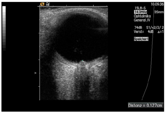
Fig. 8. Measurement of papilledema using ultrasound: disc elevation is quantified by putting the first caliper on the uppermost part of the swollen disc; the second caliper is positioned on the strongly reflecting line representing the lamina cribrosa 8

Neuro-ophthalmology Question of the Week: Fundus Autofluorescence

Question: Which of the following are hyperautofluorescent?
1. Optic Disc Drusen
2. Papilledema
3. Accumulation of lipofuscin in the retinal pigment epithelium
4. Loss of lipofuscin in the retinal pigment epithelium
5. Central serous chorioretinopathy
6. Best disease

1

Neuro-ophthalmology Question of the Week: Electroretinography

Which of the following statements are correct?
1. Full-field ERG is useful in detecting diffuse retinal disease in the setting of generalized or peripheral vision loss.
2. The ERG is invariably severely depressed by the time patients complain of visual loss.
3. In full-field ERG the responses cannot be substantially altered voluntarily.
4. The full-field ERG may be normal in minor or localized retinal disease, particularly maculopathies, even with severe visual acuity loss.
5. Multifocal ERG is extremely helpful in detecting occult focal retinal abnormalities within the macula.
6. Uncooperative patients can alter the responses on a multifocal ERG by not fixating accurately.

Neuro-ophthalmology Question of the Week: Visual Evoked Responses

Question: Which of the following are correct for visual evoked responses?
1.Abnormal responses may occur if the patient does not look at the screen, does not focus on the screen, moves the tested eye, or is tired.
2. Appropriate refraction is necessary.
3. The visual evoked response reflects the integrity of the afferent visual pathway (damage anywhere from the retina to the occipital cortex may alter the signal).
4. It is primarily a function of central visual function because such a large region of the occipital cortex near the recording electrodes is devoted to macular projections.
5. Visual evoked responses are not useful in evaluating the integrity of the visual pathway in infants and inarticulate adults.
6. Visual evoked responses cannot provide an estimate of visual acuity when stimuli of various sizes are used.

1

Neuro-ophthalmology Question of the Week: Bilateral Occipital Lobe Lesions & Visual Acuity

Question: When bilateral lesions of the retrochiasmal visual pathways produce a decrease in visual acuity which of the following are correct?
1. Such lesions produce symmetric VA loss in both eyes.
2. The VA loss from such lesions will be worse on the opposite side of the largest lesion.
3. The VA loss from such lesions will be worse on the same side of the largest lesion.
4. With the presence of retrochiasmal visual pathway lesions and different VA in the two eyes, an additional problem anterior to the chiasm must be present.

1

Neuro-ophthalmology Question of the Week: Optic Tract Defects

Question:
Which of the following are correct for a left optic tract lesion?
1. Right homonymous hemianopia
2. Left RAPD
3. Bowtie atrophy of the right optic nerve
4. Mostly temporal pallor of the left optic nerve

1

Neuro-ophthalmology Question of the Week: Ancillary Tests

Questions:
1. In the photostress test, how long does the patient look at a bright light held a few centimeters from the eye?
2. In the photostress test, what is the normal recovery time to within 1 line of best corrected visual acuity? 
3. What anatomic factor explains the RAPD with an optic tract lesion?
4. What are 5 clinical settings where OKN testing may be helpful?
5. Where is the lesion likely to be located in a patient with homonymous hemianopia and asymmetric OKN response?

1

Neuro-ophthalmology Question of the Week: Neuro-ophthalmology Coma Eye Exam

Question:
Which of the following are correct for a patient?
1. The eyes of a patient in coma will be closed.
2. Because the pathways governing ocular motility traverse the entire brainstem, brainstem lesions will most often result in abnormal eye movements, and the lesion can be localized to the midbrain, pons, or medulla.
3. The presence of a dysconjugate ocular deviation in coma (horizontal, vertical, or oblique misalignment) often indicates a cranial nerve palsy or skew deviation.
4. Roving eye movements: slow ocular conjugate deviations in random directions indicate intact ocular motility function in the brainstem.
5. Periodic alternating (“ping-pong”) gaze: slow, repetitive, rhythmic, back-and-forth, horizontal conjugate eye movements indicates intact ocular motility function in the brainstem.
6. Normal vertical and horizontal oculocephalic reflexes indicate intact ocular motility function in the brainstem.

1

Neuro-ophthalmology Question of the Week: Optokinetic Testing

Question: In which of the following conditions is optokinetic stimulus testing useful?
1. 
Infantile nystagmus
2. Patients claiming complete blindness
3. Homonymous hemianopia
4. Internuclear ophthalmoplegia
5. Dorsal midbrain syndrome
6. Optic neuritis

1

Neuro-ophthalmology Question of the Week: Optic Neuropathy or Maculopathy

Question:
Which of the following clinical tests can help differentiate between optic neuropathy and maculopathy in a patient with a reduction in visual acuity?
1.Swinging flashlight pupillary reactions
2. Amsler Grid
3. Dilation lag in darkness
4. Anisocoria
5. Photostress recovery test
6. Color vision/saturation

1

https://neuro-ophthalmology.stanford.edu/2017/02/378/