Recommended Reading – Pupillary sign of aberrant regeneration of the third nerve
Pupillary sign of aberrant regeneration of the third nerve
Olga R. Rosenvald, Simmons Lessell. Neurology. 2016; 86 (18)
VIDEO NEUROIMAGES
A 55-year-old woman presented with a third nerve palsy and impaired abduction of her right eye. MRI showed a lesion at the orbital apex extending into the cavernous sinus. A biopsy showed invasive Aspergillus fumigatus and she was treated with antifungals with only partial improvement.
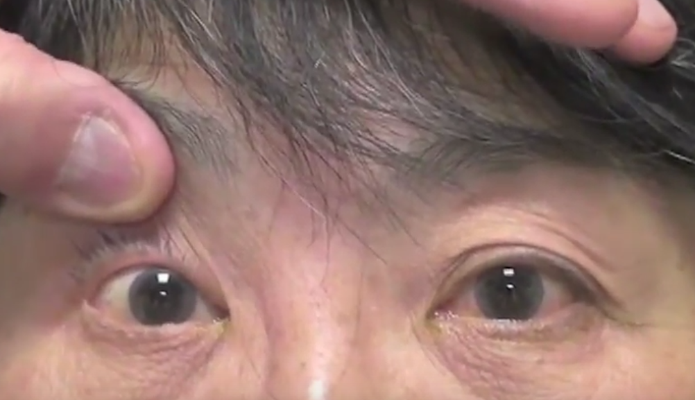
Twelve years later, she had complete ptosis, partially restricted abduction, adduction, and depression of the right eye. The right pupil, which did not constrict to light or near vision, constricted on downgaze (video on the Neurology® Web site at Neurology.org), diagnostic of aberrant regeneration,1 which occurs when regenerating axons are misdirected to muscles they were not intended, such as the iris sphincter.2
Video https://drive.google.com/open?id=1D0qDczD0jsQ7q88eUL0KzVmMhlIOqqXf
The video shows a partial cranial nerve III palsy with a pupil reactive to neither light or near vision. However, the pupil constricts on attempted downgaze.
References:
1. Czarnecki JS, Thompson HS. The iris sphincter in aberrant regeneration of the third nerve. Arch Ophthalmol 1978;96:1606–1610.
2. Sibony PA, Lessell S, Gittinger JW. Acquired oculomotor synkinesis. Surv Ophthalmol 1984;28:382–390.