Teaching NeuroImages: Kearns-Sayre syndrome
Michael T.B. Nguyen, Jonathan Micieli, Edward Margolin
RESIDENT & FELLOW SECTION. Neurology. 2019; 92 (5)
Article
A 19-year-old man presented 6 months postimplantation of permanent pacemaker for complete heart block with bilateral nonfatigable symmetric ptosis, diminished levator superioris function, and symmetric ophthalmoplegia (figure 1). Funduscopy revealed bilateral pigmentary retinopathy (figure 2). Skeletal muscle biopsy revealed presence of ragged-red fibers, consistent with Kearns-Sayre syndrome. This mitochondrial disorder is characterized by the triad of onset before age 20, chronic progressive external ophthalmoplegia, and pigmentary retinopathy. Other findings can include complete heart block, cerebellar ataxia, deafness, and endocrinopathies. CSF folate levels should be measured and supplemented if low. There is no definitive treatment but annual surveillance for comorbidities is required.1,2
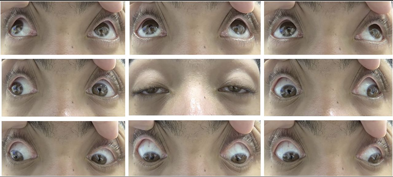
Figure 1 Ptosis and ophthalmoplegia
Photographs of eye movement demonstrate severe bilateral ptosis and mild diffuse ophthalmoplegia in all directions of attempted gaze.
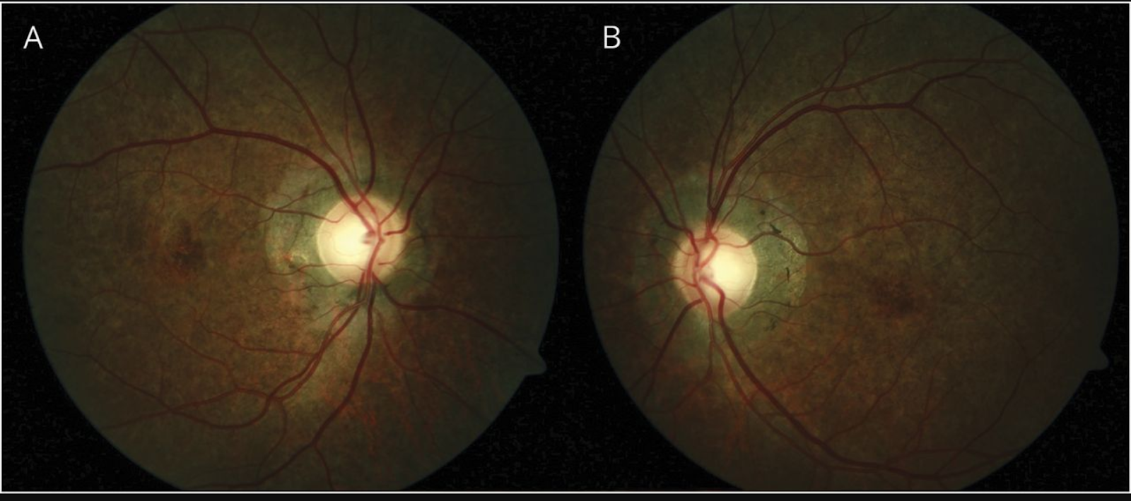
Figure 2 Pigmentary retinopathy
Fundus examination of right (A) and left eye (B) shows bilateral pigmentary retinopathy. There is diffuse depigmentation of the retinal pigment epithelium in a salt-and-pepper pattern of pigment clumping and involvement of the peripapillary zone.
References
1. Kearns TP, Sayre GP. Retinitis pigmentosa, external ophthalmoplegia, and complete heart block. Arch Ophthalmol 1958;60:280–289.
2. Shemesh A, Margolin E. Kearns Sayre syndrome. In: StatPearls [Internet]. Treasure Island, FL: StatPearls Publishing; 2018.