
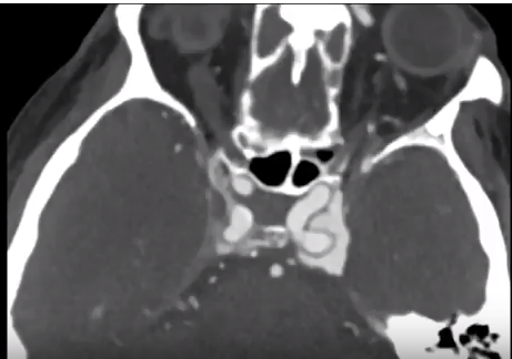
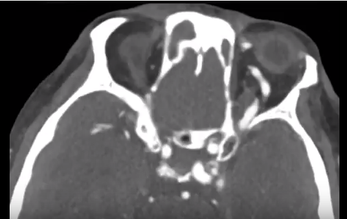
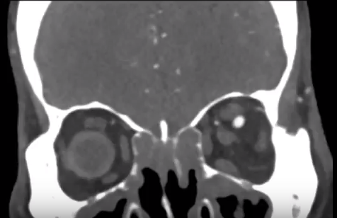
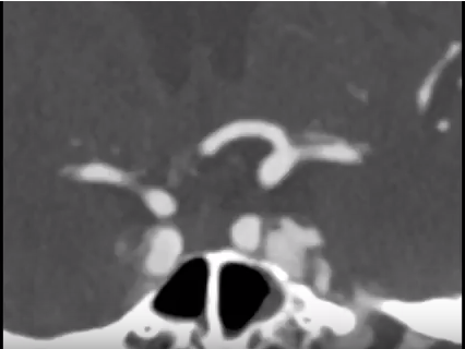
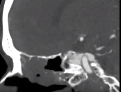
These first five CTA images of the head demonstrate filling of the left cavernous sinus in the arterial phase and asymmetric enlargement and filling of the left superior ophthalmic vein. These findings are consistent with a carotid cavernous fistula. The diagnostic angiogram was performed to evaluate the supply and drainage of the fistula. No aneurysm was identified. The fistula was supplied most prominently from the bilateral external carotid arteries and showed prominent retrograde drainage into the dilated left superior ophthalmic vein. These fistulas may present with unilateral or bilateral proptosis and chemosis and if severe may cause vision loss. This patient was treated with transvenous coil embolization and demonstrated no evidence of fistula on two month follow-up imaging.
Video https://www.youtube.com/watch?v=gkhd36dssqA
Reference
CT is us is created and maintained by The Advanced Medical Imaging Laboratory (AMIL). AMIL is a multidisciplinary team dedicated to research, education, and the advancement of patient care using medical imaging with a focus on spiral CT and 3D imaging. The AMIL is headed by Elliot K. Fishman, M.D.