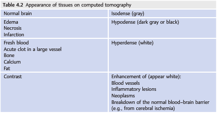
Question: On CT which of the following are isodense, hypodense, hyperdense, or enhance on contrast?
1 Acute clot in a large vessel
2 Blood vessels
3 Bone
4 Breakdown of the normal blood-brain barrier
5 Calcium
6 Edema
7 Fat
8 Fresh blood
9 Infarction
10 Inflammatory lesions
11 Necrosis
12 Neoplasms
13 Normal brain

______________________________________________________
Question with answers: On CT which of the following are isodense, hypodense, hyperdense, or enhance on contrast?
1 Acute clot in a large vessel = Hyperdense
2 Blood vessels = Enhance on contrast
3 Bone = Hyperdense
4 Breakdown of the normal blood-brain barrier = Enhance on contrast
5 Calcium = Hyperdense
6 Edema = Hypodense
7 Fat = Hyperdense
8 Fresh blood = Hyperdense
9 Infarction = Hypodense
10 Inflammatory lesions = Enhance on contrast
11 Necrosis = Hypodense
12 Neoplasms = Enhance on contrast
13 Normal brain = Isodense
Explanation1: “Computed Tomography
Because bone, calcification, fat, and blood all have unique X-ray absorption patterns, CT is a very effective technique for orbital imaging (▶Table 4.2).
Specific absorption patterns can be highlighted on CT to emphasize bone, soft tissues, or blood.
CT images are classically obtained in the axial planes. It is possible to also request images in the coronal plane by repositioning the patient. Sagittal images may be obtained by computer reformatting.
Routine studies are done at 3 or 5mm slice intervals, but it is possible to obtain 1mm slice intervals (better resolution).
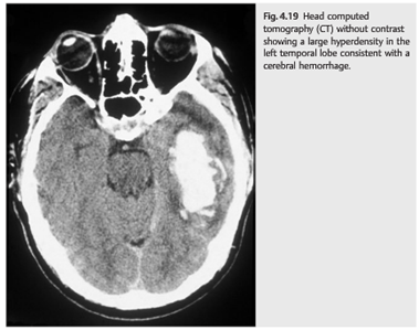
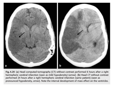
A head CT without contrast takes only a few minutes and is readily available. It is commonly performed in the emergency room and is extremely valuable in trauma patients (who may have a bone fracture or an orbital foreign body), in stroke patients (▶Fig. 4.19 and ▶Fig. 4.20), and when an acute intracranial or intraorbital hemorrhage is suspected (e.g., to detect a subarachnoid hemorrhage in a patient with an explosive headache). However, a normal head CT without contrast is insufficient in almost all other situations. It is falsely reassuring and often misses serious disorders.
The following are good indications for orbital CT:
- Orbital trauma (suspected fractures or foreign body)
- Ocular trauma to rule out a foreign body (ruptured globe)
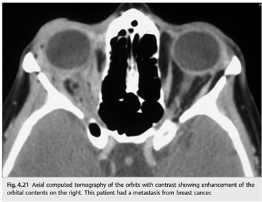
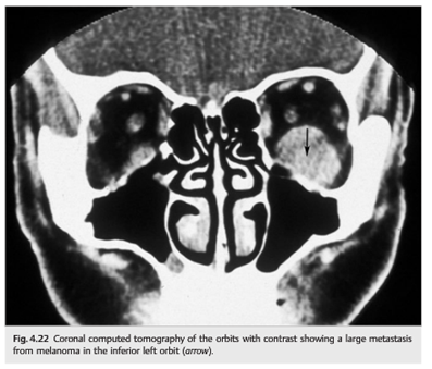
- Infectious or noninfectious orbital inflammation (▶Fig. 4.21 and ▶Fig. 4.22)
- Bone lesions (osteoma, fibrous dysplasia, suspected metastatic disease, etc.)
- Preoperative imaging for orbital disease (when imaging of the facial sinuses is very important)
- Lesions that may contain calcium (retinoblastoma, optic nerve drusen, orbital varix, meningioma, etc.)
- Lacrimal gland lesions
Reference:
1. Neuro-ophthalmology Illustrated-2nd Edition. Biousse V and Newman NJ. 2012. ThemeMore than 600 additional neuro-ophthalmology questions are freely available at http://EyeQuiz.com.
Questions prior to September 2016 are archived at http://ophthalmology.stanford.edu/blog/
After that, questions are archived at https://neuro-ophthalmology.stanford.edu
Follow https://twitter.com/NeuroOphthQandA to be notified of new neuro-ophthalmology questions of the week.
Please send feedback, questions and corrections to tcooper@stanford.edu.