Teaching NeuroImages: Ocular bruit in carotid-cavernous sinus fistula
Jeong-Yoon Choi, Seol-Hee Baek, Jin-Man Jung, Do-Young Kwon, Moon Ho Park
Neurology. August 12, 2014; 83 (7) RESIDENT AND FELLOW SECTION
ARTICLE
A 57-year-old man who had a traffic accident 1 month previously presented with left ocular pain, double vision, and left eye proptosis with ptosis and conjunctival hemorrhage. Fundus showed dilated veins with no hemorrhages or disc edema. Left ocular motility showed complete external ophthalmoplegia (figure 1). There was prominent ocular bruit in his left eye (audio file on the Neurology® Web site at Neurology.org). MRI and magnetic resonance angiography showed a dilated left superior ophthalmic vein and an extravasation into cavernous sinus (figure 2). With chemosis, ophthalmoplegia, and retro-orbital pain, the auscultation of orbital bruit can make a correct and prompt diagnosis in the patient with carotid-cavernous sinus fistula.1
Figure 1 Physical examination
(A) Left eye with conjunctival injection and ptosis.
(B) Left eye proptosis.
(C) Fundus shows dilated veins with no hemorrhages or disc edema.
(D) Ocular motility shows complete external ophthalmoplegia in left eye and partial limitation of abduction in right eye.

Figure 2 Brain MRI and magnetic resonance angiography findings
(A) magnetic resonance angiography
(B) show a dilated left superior ophthalmic vein (black arrowhead) and a extravasation into cavernous sinus (white arrow).

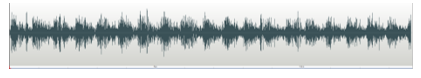
Audio. Auscultation of ocular bruit.
(audio.mp3)
It was recorded using the JABES electronic stethoscope (GS tech., Korea) and WavePad Sound Editor (NCH software, Australia).
AUTHOR CONTRIBUTIONS Dr. Choi: participated in conceptualization of the manuscript, drafted the manuscript. Dr. Baek: participated in analysis of results and conceptualization of the manuscript. Dr. Jung: selected appropriate images and revised the manuscript for intellectual content. Dr. Kwon: participated in analysis of results and revised the manuscript for intellectual content. Dr. Park: drafted the manuscript and figure legend and revised the manuscript for intellectual content.
STUDY FUNDING No targeted funding reported.
DISCLOSURE The authors report no disclosures relevant to the manuscript. Go to Neurology.org for full disclosures.
Footnotes
Go to Neurology.org for full disclosures. Funding information and disclosures deemed relevant by the authors, if any, are provided at the end of the article.
Supplemental data at Neurology.org
Download teaching slides: Neurology.org
© 2014 American Academy of Neurology
REFERENCE 1. Ling JD, Chao D, Al Zubidi N, Lee AG. Big red flags in neuro-ophthalmology. Can J Ophthalmol2013;48:3–7.