Questions:
1. True or False: The term stroke includes cerebral ischemia (transient ischemic attacks and cerebral infarctions) and cerebral hemorrhage.
2. True or False: Central retinal artery and branch retinal artery occlusions are equivalent to strokes?
3. What are 12 manifestations of carotid disease?
4. What are 4 manifestations of carotid dissection?
5. What are 5 manifestations of carotid or vertebral artery dissections?
6. Is it correct that in most cases of ocular or cerebral ischemia an evaluation for a cause of thrombophilia should be performed?
7. Is it common for multiple types of congenital thrombophilia to coexist in the patient?
8. Does the presence of marked papilledema with cerebral venous thrombosis require prompt treatment to lower the intracranial pressure?
9. What are 5 classic presenting findings of cerebral venous thrombosis?
10. What tests are used to demonstrate the presence of cerebral venous thrombosis?
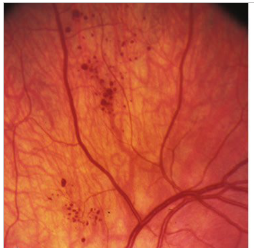
11. What retinal lesion is present in this image?
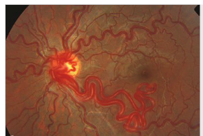
12. What retinal lesion is present in this image?
13. What syndrome do these images suggest?

14. What are the 4 classic findings of Terson syndrome?
15. What should be suspected in the presence of a patient with the following findings:
a. Headaches
b. Seizures
c. Focal neurological symptoms and signs (transient ischemic attacks,
cerebral infarctions, or cerebral hemorrhages)
d. Altered mental status?
____________________________________________________
Questions with answers:
1. True or False: The term stroke includes cerebral ischemia (transient ischemic attacks and cerebral infarctions) and cerebral hemorrhage?
True
2. True or False: Central retinal artery and branch retinal artery occlusions are equivalent to strokes?
True
3. What are 12 manifestations of carotid disease?
1. Asymptomatic retinal emboli
2. Transient monocular visual loss
3. Central or branch retinal artery occlusion
4. Ophthalmic artery occlusion
5. Episcleral artery dilation
6. Venous stasis retinopathy
7. Ocular ischemic syndrome
8. Ischemic optic neuropathy
9. Optic nerve compression
10. Horner syndrome
11. Ocular motor nerve paresis
12. Referred pain.
All of these may be a manifestation of carotid disease, but ocular motor nerve paresis, ischemic optic neuropathy, and ocular motor nerve paresis are rare.
4. What are 4 manifestations of carotid dissection?
1. Horner syndrome
2. Orbital pain
3. Face pain
4. Head pain.
These patients are at risk for cerebral infarction and should be evaluated and treated emergently.
5. What are 4 features of carotid or vertebral artery dissections?
1. They may occur spontaneously.
2. They may occur after cervical trauma (car accident, strangulation, chiropractic manipulation).
3. Pain is often present immediately after the trauma.
4. There is often a symptom-free interval of a few days between the trauma and the first sign of the dissection.
6. Is it correct that in most cases of ocular or cerebral ischemia an evaluation for a cause of thrombophilia should be performed?
Yes. There are more than 10 congenital and 12 acquired risk factors for thrombophilia. Referral to a hematologist for evaluation is recommended. The cause of thrombophilia is more likely to be found in these situations:
1. In younger patients.
2. When no other obvious risk factor for cerebral or ocular ischemia is present.
3. When a family history of thrombophilia or recurrent thrombosis is present.
4. When there is a prior history of thrombosis.
5. When there have been recurrent, unexpected episodes of thrombosis.
6. When there has been venous thrombosis at unusual sites (e.g., cerebral venous thrombosis).
7. Is it common for multiple types of congenital thrombophilia to coexist in the patient?
Yes, multiple congenital thrombophilias often coexist in the same patient; therefore, all patients at risk should be screened for all types of thrombophilia. Referral to a hematologist for evaluation is recommended.
8. What are 5 classic presenting findings of cerebral venous thrombosis?
1. Signs of raised intracranial pressure (headache, papilledema, sixth nerve palsy)
2. Seizures
3. Altered mental status
4. Neurologic deficit (hemiparesis, aphasia based on the location of cerebral infarctions)
5. Deficits on alternating sides or occurring bilaterally (unlike in cerebral arterial ischemia).
Urgent treatment is necessary to prevent multiple cerebral venous infarctions and death.
9. Does the presence of marked papilledema with cerebral venous thrombosis require prompt treatment to lower the intracranial pressure?
Permanent visual loss from papilledema is a classic complication of cerebral venous thrombosis. Early treatment of intracranial hypertension is necessary. When possible, a lumbar puncture should be performed prior to anticoagulation to reduce the intracranial pressure and help preserve vision.
10. What tests are used to demonstrate the presence of cerebral venous thrombosis?
MRI & MRV.
11. What retinal lesion is present in this image?

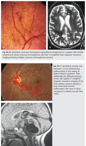
Cavernous hemangioma
12. What retinal lesion is present in this image?

A true AVM.
13. What syndrome do these images suggest?
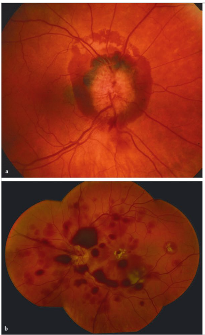
Terson Syndrome – Intraocular hemorrhage associated with subarachnoid hemorrhage. Images show optic nerve edema, peripapillary hemorrhages, and vitreous hemorrhage associated with aneurysmal rupture and subarachnoid hemorrhage.
14. What are the 4 classic findings of Terson syndrome?
1. Optic nerve head edema, often with hemorrhages
2. Retinal hemorrhages
3. Subhyaloid hemorrhages
4. Vitreous hemorrhages
15. What should be suspected in the presence of a patient with the following 4 findings:
a. Headaches
b. Seizures
c. Focal neurological symptoms and signs (transient ischemic attacks, cerebral infarctions, or cerebral hemorrhages)
d. Altered mental status?
Cerebral vasculitis – The presence of retinal vasculitis is rare and usually suggests a systemic vasculitis.
____________________________________________________
The information below is from: Neuro-ophthalmology Illustrated-2nd Edition. Biousse V and Newman NJ. 2012. Thieme
20 Disorders Commonly Encountered in Neuro-ophthalmology
Some neurologic or systemic disorders are commonly encountered in neuro-ophthalmology. Patients with common neurologic disorders such as multiple sclerosis, strokes, traumatic brain injuries, or brain tumors may have visual loss or diplopia. However, it is important to realize that these patients often can also have routine ocular disorders(such as refractive errors, corneal surface disease, or cataracts) that can be easily treated. In addition, even patients with neurologic disorders can have urgent acute ocular disorders such as retinal detachment, retinal vascular occlusions, or intraocular hypertension.
Pearls
Neurologists evaluating a known neurologic patient with new-onset visual loss should not automatically assume that visual loss is related to the neurologic disease. They should obtain an ophthalmic consultation prior to making management and therapeutic decisions.
Some disorders, such as pituitary tumors, occipital lesions, intracranial aneurysms, and raised intracranial pressure, commonly affect the visual pathways. Systematic neuro-ophthalmologic follow-up with careful examination of visual function (including repeat visual fields) can document worsening of the intracranial process.
A few diseases have pathognomonic ocular findings that facilitate early diagnosis. These patients are often sent to neuro-ophthalmology for “screening.” This is often the case when the diagnoses of Wilson disease, Whipple disease, or neurofibromatosis type 1 are being considered, or when it is necessary to determine if an incidental finding (such as Chiari malformation or an intracranial aneurysm) is symptomatic and, therefore, should be treated.
20.1 Cerebrovascular Disease
The term stroke includes cerebral ischemia (transient ischemic attacks and cerebral infarctions) and cerebral hemorrhage. A stroke is suspected when a patient presents with acute neurologic symptoms and signs (acute usually means that the mechanism is vascular). Stroke patients often have neuro-ophthalmic complaints, including visual loss, visual field defects, and diplopia. Retinal vascular disorders are equivalent to strokes involving various ocular vascular territories.
Once the diagnosis of stroke is suspected, the first step is to determine whether the vascular event is ischemic or hemorrhagic. In the eye, this can be done by looking at the fundus; for the brain, it requires neuroimaging, usually a head computed tomographic (CT) scan without contrast (▶Fig. 20.1 and ▶Fig. 20.2)

Questions to be answered in patients with suspected cerebrovascular disease (and retinal vascular diseases) include the following:
1. Is it a vascular event?
2. Where in the brain or the eye has the vascular event occurred (parenchymal and vascular topography)?
3. What type of vascular event is it (pathology)?
4. What has caused the vascular event (mechanism)?
5. What are the consequences of the vascular event (impairments, disabilities, and handicap)?
6. What other medical problems coexist?
20.1.1 Cerebral Infarctions
Once a stroke is confirmed and the mechanism (ischemic vs. hemorrhagic) is determined, the cause should be clarified to offer the best secondary prevention to the patient (▶Fig. 20.3).

Causes of Cerebral Infarction (see ▶Fig. 20.3)
Four main mechanisms can result in cerebral ischemia.
● Thrombosis of a vessel○ Large-vessel or macrovascular (arterial) disease○ Small-vessel or microvascular (arterial) disease
● Emboli
○ Cardiac source of emboli
○ Artery to artery
● Hypoperfusion
● Venous thrombosis
Ocular Manifestations of Carotid Disease
Carotid disease (mostly internal carotid artery) often presents with ocular symptoms and signs.
● Asymptomatic retinal emboli
● Transient monocular visual loss
● Central or branch retinal artery occlusion
● Ophthalmic artery occlusion
● Episcleral artery dilation
● Venous stasis retinopathy
● Ocular ischemic syndrome
● Ischemic optic neuropathy (rare)
● Optic nerve compression (rare)
● Horner syndrome
● Ocular motor nerve paresis (rare)
● Referred pain
Differential Diagnosis of Carotid Artery Disease
The carotid artery can be affected by the following diseases:
● Arterial wall
○ Atheroma
○ Dissection
○ Fibromuscular dysplasia
○ Arteritis
– Infectious
– Noninfectious (Takayasu, giant cell arteritis)
○ Trauma
○ External radiation
○ Tumors (carotid glomus)
● External compression
○ Tumors
○ Trauma
● Blood flow
○ Coagulation disorders
○ Emboli (heart, artery to artery)
Cervical Artery Dissections
Dissections of the internal carotid artery commonly present with an ipsilateral acute Horner syndrome associated with orbital, face, or head pain. These patients are at risk for a cerebral infarction and should be evaluated and treated emergently (▶Fig. 20.4and ▶Fig. 20.5).


Dissections involve the extracranial carotid or vertebral arteries more often than the intracranial arteries. They may occur spontaneously or after cervical trauma (car accident, strangulation, chiropractic manipulation). There is often a symptom-free interval of a few days between the trauma and the first sign of the dissection. Pain is often present immediately after the trauma.
Cardiac Sources of Embolism and Embolic Risk
The risk of cerebral emboli in cardiac disease is classically defined as “high” (needing urgent treatment) and “low or uncertain” (often not directly responsible for the cerebral infection)
High Risk
● Atrial
○ Atrial fibrillation
○ Sustained atrial flutter
○ Sick sinus syndrome
○ Left atrial thrombus
○ Left atrial appendage thrombus
○ Left atrial myxoma
● Valvular disease
○ Mitral stenosis
○ Prosthetic valves
– Mechanical
– Bioprosthetic
○ Endocarditis
– Infective
– Noninfective
a. Marantic (Non-bacterial thrombotic endocarditis (NBTE) is a form of endocarditis in which small sterile vegetations are deposited on the valve leaflets. Formerly known as marantic endocarditis)
b. Liebman Sachs (systemic lupus erythematosus; antiphospholipid antibodies)
○ Ventricular
– Recent anterior myocardial infarction
– Left ventricular thrombus
– Left ventricular myxoma
– Dilated cardiomyopathy
○ Iatrogenic
– Cardiac catheterization
– Cardiac surgery
Low or Uncertain Risk
● Atrial
○ Patent foramen ovale
○ Atrial septal aneurysm
○ Spontaneous echo contrast on transesophageal echocardiogram (TEE)
● Valvular disease
○ Mitral annulus calcification
○ Mitral valve prolapse
○ Calcified aortic stenosis
○ Fibroelastoma
○ Giant Lambl excrescences
● Ventricular
○ Akinetic/dyskinetic ventricular wall segment
○ Subaortic hypertrophic cardiomyopathy
○ Congestive heart failure
Classification of Small Vessel Disease
Cerebral infarctions can be related to occlusion of a large intracranial vessel (such as posterior cerebral artery or middle cerebral artery), or can be related to diseases affecting small intracranial vessels. Small vessel diseases include abnormalities in the vessel content and vessel wall abnormalities.
20.1.2 Abnormalities in the Vessel Content
● Hypercoagulable states
20.1.3 Vessel Wall Abnormalities (Veins and Arteries)
● Acute
○ Vasculitis
○ Noninflammatory vasculopathies
● Chronic
○ Arteriolar sclerosis (“lacuna” due to hypertension)
○ Cerebral amyloid angiopathy
○ Cerebral autosomal dominant arteriopathy with subcortical infarcts and leukoencephalopathy (CADASIL)
○ Mitochondrial encephalopathy with lactic acidosis and stroke-like episodes (MELAS)
Risk Factors for Ischemic Stroke
Vascular risk factors should be evaluated in all patients with cerebral or ocular ischemia. Aggressive treatment of modifiable risk factors is essential in secondary prevention.
Risk factors for ischemic stroke include the following:
● Nonmodifiable
○ Age
○ Gender (men>women)
○ Ethnicity (African Americans and Hispanics>Caucasians)
○ Heredity
○ Migraine
● Modifiable
○ Elevated blood pressure
○ Cardiovascular disease
○ Diabetes
○ Hyperlipidemia
○ Cigarette smoking
○ Alcohol abuse
○ Obesity
○ Sedentary lifestyle
○ Obstructive sleep apnea
○ Asymptomatic carotid stenosis
○ Hyperhomocysteinemia
○ Chronic infection
○ Oral contraceptives
Laboratory and Diagnostic Tests Recommended for Patients with Suspected Stroke
Some tests are systematically obtained in the emergency department in patients with suspected strokes. Other tests are obtained based on the patient’s characteristics and risk factors.
These tests include the following (* tests are indicated only for certain patients, depending on stroke type and clinical setting):
● Laboratory
○ Complete Blood count
○ Platelet count
○ Blood glucose level
○ Serum electrolytes, including magnesium and calcium
○ Serum creatinine level
○ Prothrombin time and activated partial prothrombin time, international normalized ratio
○ Urinalysis (may detect occult blood indicating embolic events in the kidney)
○ Hepatic function tests*
○ Toxicology screen*
○ Blood alcohol determination*
○ Pregnancy test*
● Other tests
○ Electrocardiogram (or cardiac monitoring)
○ Chest X-ray (helpful in assessing cardiac disease and aspiration pneumonia)
○ Brain CT or magnetic resonance imaging (MRI)—often with computed tomographic angiography (CTA) or magnetic resonance angiography (MRA) of head and neck
○ Carotid duplex ultrasound* (in anterior circulation infarctions when CTA or MRA are not performed immediately)
○ Holter monitor*
○ Transthoracic or transesophageal echocardiogram*
○ Lumbar puncture*
Hypercoagulable States
Hypercoagulable states can produce a cerebral or retinal infarction by occluding an artery. Many of these factors are congenital (thrombophilia), and the thrombotic episode is triggered by an acquired factor. For example, a woman born with congenital activated protein C resistance may have a normal childhood and may develop a cerebral venous thrombosis only when she starts an oral contraceptive pill or when she is pregnant.
Hypercoagulable states are only rarely responsible for cerebral or ocular arterial ischemia. The workup should be obtained only in specific situations, such as the following:
● Younger patients
● No obvious risk factor for cerebral or ocular ischemia
● Family history of thrombophilia, or recurrent thrombosis
● Prior history of thrombosis
● Recurrent, unexplained episodes of thrombosis
● Venous thrombosis at unusual sites (e.g., cerebral venous thrombosis)
Risk Factors for Thrombosis
● Congenital factors
○ Protein C defect/deficiency
○ Protein S defect/deficiency
○ Antithrombin III deficiency
○ Activated protein C resistance (factor V Leiden)
○ Prothrombin gene (factor II 20210A) mutation
○ Heparin cofactor II deficiency
○ Dysfibrinogenemi
○ Plasminogen activator inhibitor (PAI-1) gene polymorphism
○ Congenital plasminogen deficiency
○ Thrombomodulin gene mutation
○ Sickle cell disease
○ Platelet defects
● Acquired factors
○ Antiphospholipid syndrome
○ Myeloproliferative disorder○ Paroxysmal nocturnal hemoglobinuria
○ Thrombotic thrombocytopenic purpura
○ Disseminated intravascular coagulation
○ Malignancy
○ Sepsis
○ Hyperviscosity syndrome
○ Trauma
○ Immobilization
○ Surgery
○ Pregnancy
○ Oral contraceptives
○ Heparin-induced thrombocytopenia
● Combined risk (both acquired and genetic factors)
○ Hyperhomocysteinemia
○ Elevated factor VIII levels
○ Elevated fibrinogen levels
Pearls
Multiple congenital thrombophilia often coexist in the same patient; therefore, all patients at risk should be screened for all types of thrombophilia.
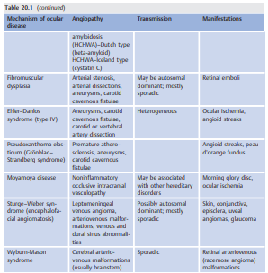
Angiopathies of the Central Nervous System Associated with Ocular Manifestations
▶ Table 20.1 lists angiopathies of the central nervous system associated with ocular manifestations.

20.1.4 Cerebral Venous Thrombosis
Because a large part of the cerebrospinal fluid (CSF) drains into the venous sinuses and the internal jugular veins (▶Fig. 20.6 and ▶Fig. 20.7), thrombosis of an intracranial venous sinus usually results in raised intracranial pressure with headaches and papilledema. Ultimately, the thrombus may extend to the deep cerebral veins and the cortical veins, resulting in acute cerebral venous infarctions and hemorrhages.


The cortical veins empty into the dural venous sinuses, which have an anteroposterior drainage into the transverse sinuses and the jugular veins. Occlusion of a sinus usually results in reversal of the flow in some veins, producing specific clinical manifestations based on the anatomical location of the thrombosed sinus (e.g., when the cavernous sinus is thrombosed, the orbital veins drain anteriorly instead of posteriorly, and there is orbital congestion with proptosis). In most cases, the CSF drainage is compromised, and there are symptoms and signs of raised intracranial pressure.
Dilation and thrombosis of the cortical veins produce catastrophic venous infarctions that are often hemorrhagic.
There are multiple veins draining the cerebellum and the brainstem (▶Fig. 20.8).

Thrombosis of some veins may result in dilation of these veins and compression or ischemia of the adjacent cranial nerves. This explains why petrosal sinus thrombosis can produce multiple cranial nerve palsies such as sixth, fifth, seventh, and third nerve palsies. Isolated diplopia with pain may rarely be the first sign of cerebral venous thrombosis.
Classic clinical presentations of cerebral venous thrombosis include the following:
● Raised intracranial pressure (headache, papilledema, sixth nerve palsy)
● Seizures
● Altered mental status
● Neurologic deficit (hemiparesis, aphasia based on the location of cerebral infarctions)
● Deficits on alternating sides or occurring bilaterally (unlike in cerebral arterial ischemia)
Urgent treatment is necessary to prevent multiple cerebral venous infarctions and death.
Pearls
Permanent visual loss from papilledema is a classic complication of cerebral venous thrombosis. Early treatment of intracranial hypertension is necessary. When possible, a lumbar puncture should be performed prior to anticoagulation to reduce the intracranial pressure and help preserve vision.
Diagnosis of Cerebral Venous Thrombosis
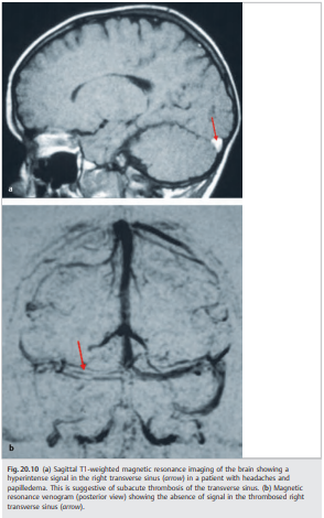
MRI and magnetic resonance venography (MRV) usually allow very good noninvasive visualization of the intracranial venous sinuses (▶Fig. 20.9 and ▶Fig. 20.10). Artifacts are common, and CT venography often complements these tests. A catheter cerebral venogram is only rarely required.

20.1.5 Intracranial Hemorrhage
Intracranial hemorrhages are classified as follows (▶Fig. 20.11):

● Epidural hemorrhage (between the skull and meninges): Usually results from skull fracture (temporal bone with rupture of the middle meningeal artery). There can be rapid expansion of the hematoma with uncal herniation, ipsilateral third nerve palsy, and death if the hematoma is not drained emergently.
● Subdural hemorrhage (between the dura and the subarachnoid space): Usually results from mild head trauma or may be spontaneous (rupture of the bridging veins).Particularly common in the elderly. There is relatively slow expansion of the hematoma with headaches and mass effect on the adjacent cerebral hemisphere. Visual field defects are common. Subdural hematoma can be subacute (over days) or chronic (over months).
● Subarachnoid hemorrhage (in the subarachnoid space): Usually results from aneurysmal rupture, or may be spontaneous or from head trauma. The blood in the subarachnoid space can produce arterial spasm with cerebral ischemia, or it may block CSF passage and cause obstructive hydrocephalus.
● Intraparenchymal hemorrhage (▶Fig. 20.12 and ▶Fig. 20.13): Intraparenchymal Hemorrhages usually result from bleeding of the small perforating arteries and most often involve the basal ganglia. Superficial intracerebral hemorrhages are often associated with subarachnoid hemorrhage from aneurysmal or arteriovenous malformation rupture.

Risk Factors for Intraparenchymal Hemorrhage
Cerebral hemorrhages may result from the following:
● Arterial hypertension
● Vascular malformations
○ Arteriovenous malformations
○ Cavernous hemangiomas
○ Aneurysms
● Cerebral amyloid angiopathy
● Brain tumor/metastases
● Bleeding disorders
○ Coagulopathies
○ Thrombocytopenia
○ Anticoagulants
○ Thrombolytic treatment
● Head trauma\
● Vasculitis
● Endocarditis
● Cerebral venous thrombosis
● Drugs (sympathomimetic agents)
● Alcohol use
● Low cholesterol
When evaluating a patient with an acute intraparenchymal hemorrhage, it is important to determine the source of the hemorrhage. It is sometimes impossible acutely because the hemorrhage may hide an underlying lesion. Repeat brain imaging a few weeks later(once the blood has partially resolved) sometimes allows visualization of a cavernous hemangioma or a mass (▶Fig. 20.14 and ▶Fig. 20.15).

Funduscopic examination is sometimes useful by revealing retinal vascular malformations (▶Fig. 20.16 and ▶Fig. 20.17).
Subarachnoid Hemorrhage
Bleeding in the subarachnoid space (▶Fig. 20.18) is usually revealed by an acute, explosive headache. There may be a third nerve palsy if the subarachnoid hemorrhage is related to rupture of an aneurysm of the posterior communicating artery (▶Fig. 20.19).


The prognosis of subarachnoid hemorrhage is poor. Immediate diagnosis and treatment are essential. Subarachnoid hemorrhage should be suspected in all patients presenting with a very severe headache. Other neurologic symptoms and signs depend on the cause of the subarachnoid hemorrhage and the location of the aneurysm (is related to an aneurysmal rupture). The most common complications are vasospasm with cerebral infarction and obstructive hydrocephalus.
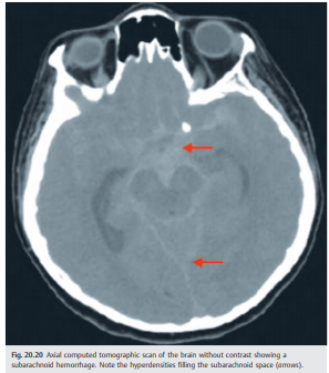
Visual fields often reveal a contralateral homonymous defect when the vascular malformation involves the retrochiasmal visual pathways (▶Fig. 20.20).
Etiologies of Subarachnoid Hemorrhage
● Aneurysm rupture
● Vascular malformation bleeding
● Bleeding diathesis
● Trauma
● Drug (cocaine, methamphetamine)
● Amyloid angiopathy
● Hypertension
● Brain tumors
● Spinal lesions
○ Aneurysms
○ Arteriovenous malformations
○ Tumors
Terson Syndrome
Subarachnoid hemorrhage produces a very acute increase in intracranial pressure, sometimes associated with Terson syndrome (retinal and vitreous hemorrhages)(▶Fig. 20.21 and ▶Fig. 20.22). Terson syndrome is a classic cause of unilateral or bilateral visual loss in patients with subarachnoid hemorrhage. Because these patients are often unconscious, the diagnosis of Terson syndrome is typically delayed unless funduscopic examination is systematically performed.
Classic findings include the following:
● Optic nerve head edema, often with hemorrhages
● Retinal hemorrhages
● Subhyaloid hemorrhages
● Vitreous hemorrhages

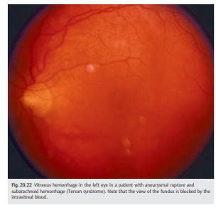
The intraocular hemorrhages likely result from acute venous pressure secondary to the subarachnoid hemorrhages; they do not result from diffusion of the blood from the subarachnoid space into the eye. In many cases, the hemorrhages resolve spontaneously over a few weeks or months. Macular hemorrhages may result in permanent visual loss. Persistent vitreous hemorrhage may require a vitrectomy for removal of the blood. Traction retinal detachment may develop. Intracranial Aneurysms Intracranial aneurysms represent the most common cause of subarachnoid hemorrhage. This is why catheter angiography is always performed immediately when a subarachnoid hemorrhage is diagnosed: early treatment of the ruptured aneurysm allows prevention of complications and rebleeding. All intracranial vessels are examined because about 20% of patients have more than one intracranial aneurysm.
Intracranial aneurysms (▶Table 20.2, ▶Fig. 20.23) may manifest in various ways:
● May be asymptomatic, found on imaging performed for another reason
● Compression of adjacent structures (mass effect of the aneurysm)
● Distal emboli from the sac of the aneurysm
● Rupture with devastating subarachnoid hemorrhage


20.2 Vasculitis (Angiitis)
Cerebral vasculitis may occur primarily (without any systemic manifestations) or maybe secondary to systemic vasculitis. The terms vasculitis and angiitis imply that there is inflammation in or around the blood vessels. It is a pathological term and the diagnosis of cerebral angiitis or vasculitis should be made only after it is confirmed by cerebral and leptomeningeal biopsy. All other cases should be called cerebral vasculopathy.
20.2.1 Patient Evaluation
Classic clinical presentations include the following:
● Headache
● Seizures
● Focal neurological symptoms and signs (transient ischemic attacks, cerebral infarcts, or cerebral hemorrhages)
● Altered mental status
● Retinal vasculitis (rare and usually suggests a systemic vasculitis)
The patient’s condition often worsens rapidly. Workup and treatment often need to be performed emergently.
Confirmation of the diagnosis involves the following steps:
● Brain MRI with contrast and diffusion-weighted imaging (▶Fig. 20.24a)
○ Increased T2 signals involving the white and gray matter (often small and multiple)
○ Cerebral infarction, cerebral hemorrhage, or diffuse white matter changes

● Electroencephalogram: often abnormal
● Lumbar puncture: often abnormal (lymphocytic meningitis)
● Vascular imaging demonstrates irregularity of the intracranial arteries
○ MRA and CTA are poorly sensitive because most cerebral angiitis involves the small intracranial vessels, which are not well visualized on MRA and CTA.
○ Catheter angiography is necessary in most cases (▶Fig. 20.24b).
● Cerebral and leptomeningeal biopsy confirms the diagnosis.
The diagnosis of cerebral angiitis should be made only after it is confirmed by cerebral and leptomeningeal biopsy.
20.2.2 Classification
Central nervous system angiitis is classified as follows:
● Cerebral angiitis associated with systemic diseases
○ Systemic vasculitides
○ Connective tissue diseases
● Cerebral angiitis associated with infections
○ Bacterial
– Bacterial meningitis
– Bacterial endocarditis
– Lyme disease
– Chlamydia pneumonia
– Mycoplasma pneumonia
– Tuberculosis
– Syphilis
– Cat scratch disease (Bartonella henselae)
○ Viral
– Herpes zoster and herpes simplex
– Cytomegalovirus
– Human immunodeficiency virus (HIV)
○ Parasitic
– Toxoplasmosis
– Cysticercosis
○ Fungal
– Aspergillosis
– Coccidioidomycosis
– Histoplasmosis
– Cryptococcosis
– Mucormycosis
● Cerebral angiitis associated with neoplasm
○ Carcinomatous meningitis
○ Lymphoma
● Primary angiitis
○ Primary granulomatous angiitis
○ Benign cerebral angiitis
○ Postpartum benign cerebral angiitis
Certain conditions mimic cerebral angiitis:
● Noninflammatory vasculopathies
○ Benign cerebral angiopathy
○ Reversible cerebral vasoconstriction syndrome
○ Cholesterol emboli
○ Susac syndrome
○ Cerebral autosomal dominant arteriopathy with subcortical infarcts and leukoencephalopathy (CADASIL)
○ Hereditary endotheliopathy with retinopathy, nephropathy, and stroke (HERNS)
○ Vasospasm (subarachnoid hemorrhage, pheochromocytoma, malignant systemic hypertension, ergotism, amphetamines, nasal spray vasoconstrictor)
○ Intracranial dissection
○ Fibromuscular dysplasia
○ Moyamoya disease
○ Sneddon syndrome
● Vasculopathies associated with drugs
○ Vasoconstrictors
○ Amphetamines
○ Cocaine
● Coagulopathies
○ Antiphospholipid antibody syndrome
○ Disseminated intravascular coagulation
○ Thrombotic thrombocytopenic purpura
○ Sickle cell disease
○ Hyperviscosity syndrome
● Cardiac emboli
● Severe infections
○ Meningococcemia
○ Hemorrhagic fever
○ Malaria
○ Leptospirosis
● Neoplasia
○ Central nervous system lymphoma
○ Endovascular lymphoma
● Metabolic diseases
○ Fabry disease
○ Cerebrotendinous xanthomatosis
○ MELAS
Reference: 1. Neuro-ophthalmology Illustrated-2nd Edition. Biousse V and Newman NJ. 2012. Thieme
These questions are archived at https://neuro-ophthalmology.stanford.edu
Follow https://twitter.com/NeuroOphthQandA to be notified of new neuro-ophthalmology questions of the week.
Please send feedback, questions, and corrections to tcooper@stanford.edu.