Questions:
1. What are the characteristics of physiologic nystagmus?
2. How is pathologic nystagmus characterized?
3. Why is it important to differentiate peripheral from central nystagmus? 4. How is peripheral nystagmus different from central nystagmus?
5. What are the findings in jerk nystagmus?
6. What are the findings in pendular nystagmus?
7. What are the characteristics of acquired pendular nystagmus?
8. What are the characteristics of infantile (congenital) pendular nystagmus?
9. What is latent nystagmus?
10. What is spasmus nutans?
11. What is infantile monocular pendular nystagmus?
12. What is acquired gaze-evoked nystagmus?
____________________________________________________
Questions with answers:
1. What are the characteristics of physiologic nystagmus?
Physiologic nystagmus or rapid gaze-evoked nystagmus is present only in extremes of horizontal gaze and dampens within seconds. It resolves when the eyes are in a slightly less eccentric position.
2. How is pathologic nystagmus characterized?
Pathologic nystagmus is characterized as jerk or pendular, and infantile (congenital) or acquired.
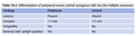
3. Why is it important to differentiate peripheral from central nystagmus?
Central nystagmus indicates a problem in the central nervous system while peripheral nystagmus indicates a problem in the peripheral vestibular system.
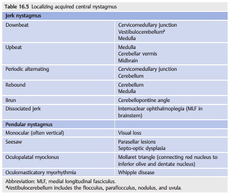
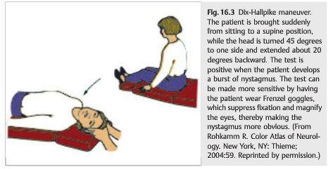
4. How is peripheral nystagmus different from central nystagmus?
Differentiation of peripheral versus central nystagmus can be determined with the Dix–Hallpike maneuver. The patient is brought suddenly from sitting to a supine position, while the head is turned 45 degrees to one side and extended about 20 degrees backward. The test is positive when the patient develops a burst of nystagmus indicating peripheral nystagmus.
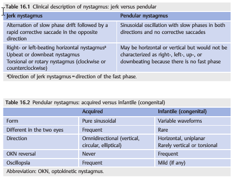
5. What are the findings in jerk nystagmus?
In jerk nystagmus, there is an alternation of slow phase drift and the following rapid corrective saccade in the opposite direction. It is characterized by the direction of the fast phase.
6. What are the findings in pendular nystagmus?
1. Sinusoidal oscillation with slow phases in both directions and no corrective saccades.
2. May be horizontal or vertical but would not be characterized as right-, left-, up-, or down beating because there is no fast phase.
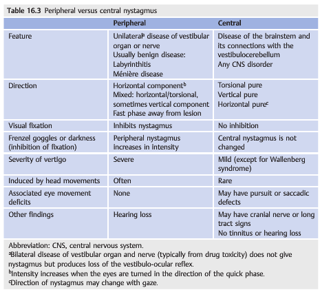
7. What are the characteristics of acquired pendular nystagmus?
1. Form=Pure sinusoidal oscillations
2. Different in the two eyes=Frequent
3. Direction=Omnidirectional (vertical, circular, elliptical)
4. OKN reversal=Never
5. Oscillopsia=Frequent
8. What are the characteristics of infantile (congenital) pendular nystagmus?
1. Form=Variable waveforms
2. Different in the two eyes=Rare
3. Direction=Horizontal, uniplanar, rarely vertical or torsional
4. OKN reversal=Frequent
5. Oscillopsia=Mild (if any)
6. Vertical OKN intact=good vision
9. What is latent nystagmus?
This is a variant of infantile nystagmus that is not evident during binocular fixation but appears when either eye is covered (uncovered eye beats away from the covered eye). It is often seen in infantile esotropia (most common), often with amblyopia, and with any lesion disrupting binocular development in the first 6 months of life. It is also common in Down syndrome. Manifest-latent nystagmus is continually present but worsens when one eye is covered.
10. What is spasmus nutans?
Spasmus nutans involves a triad of symptoms:
1. Very asymmetric and occasionally monocular nystagmus with rapid pendular eye movements,
2. Head nodding,
3. Torticollis (head tilt or head turn)
Onset is usually in the first year of life with the nystagmus typically lasting for several months. The condition is usually benign with no neurologic abnormalities. Neuroimaging is recommended as anterior visual pathway gliomas may mimic spasmus nutans. Ophthalmologic evaluation and possibly electroretinogram are recommended as retinal disorders causing visual loss may mimic spasmus nutans.
11. What is infantile monocular pendular nystagmus?
This is usually due to visual loss (often optic neuropathy or chiasmal glioma). In cases of bilateral visual loss, there is bilateral nystagmus, with greater nystagmus in the eye with the worst vision.
12. What is acquired gaze-evoked nystagmus?
Gaze-evoked is the most common type of nystagmus. It is absent in the primary position and is not visually disabling. It is a jerk nystagmus that beats in the direction of gaze. It results from impairment in eccentric gaze-holding mechanisms, such as from sedative medications/anticonvulsants, brainstem and cerebellar lesions (central vestibular dysfunction).
____________________________________________________
From: 2003 Clinical Pathways in Neuro-ophthalmology 2nd Edition – Lee & Brazis

The information below is from: Neuro-ophthalmology Illustrated-2nd Edition. Biousse V and Newman NJ. 2012. Thieme
16 Nystagmus and Other Ocular Oscillations
Nystagmus is a rhythmic, repetitive oscillation of the eyes, initiated by a slow eye movement that drives the eye o target, followed by a fast movement that is corrective(jerk nystagmus) or another slow eye movement in the opposite direction (pendular nystagmus). Saccadic intrusions (opsoclonus and flutter) are abnormal rapid eye movements (saccades) that have no slow phase. All such eye movements disrupt fixation and may interfere with vision.
16.1 Nystagmus
Nystagmus may occur physiologically in response to an environmental stimulus or change in body position. It is also seen with diseases of the central nervous system or peripheral vestibular system and in some cases of visual loss. Physiologic nystagmus or rapid gaze-evoked nystagmus is present only in extremes of horizontal gaze and dampens within seconds. It resolves when the eyes are in a slightly less eccentric position. Pathologic nystagmus is characterized as jerk or pendular, and infantile (congenital)or acquired. In patients presenting with nystagmus and vertigo, it is essential to differentiate peripheral vestibular nystagmus from central nystagmus (▶Table 16.1, ▶Table 16.2, ▶Table 16.3,▶Table 16.4, ▶Table 16.5).
Most patients with nystagmus complain of oscillopsia (oscillating vision with illusion that objects are moving), and in most cases, nystagmus can be recognized clinically without eye movement recording. However, eye movement recording allows far more accurate characterization of the nystagmus by analyzing the slow phase (velocity, amplitude, and frequency) (▶Fig. 16.1 and ▶Fig.6.2) show the waveforms of horizontal jerk and pendular nystagmus.

16.1.1 Patient Evaluation
The goals of the evaluation are to decide whether there is a central or peripheral pattern of nystagmus and to determine if localization is possible based on the findings(▶Table 16.3 and ▶Table 16.4).Symptoms include oscillopsia (absent in congenital nystagmus), decreased acuity, nausea or vomiting, and vertigo.
There may be coexisting neurologic deficits.
The examination (in primary position as well as all positions of gaze) differentiates jerk from pendular nystagmus. If the finding is jerk nystagmus, look for the direction of the fast phase—watch for a few minutes, as nystagmus may occasionally alternate directions. Look for coexisting head oscillations or head turns, the effect of convergence on nystagmus, the presence of a null point (eye position where nystagmus is least prominent), and subtle nystagmus or vestibular nystagmus that is suppressed by fixation. The last can be assessed by performing ophthalmoscopy in one eye while the patient fixates at distance, then covering the fixating eye. Nystagmus may then be viewed through the ophthalmoscope (the fast phase direction is the opposite of what it appears through the direct ophthalmoscope). Frenzel goggles may be used to assess nystagmus in the absence of fixation. Electronystagmography (ENG) is another method of identifying nystagmus not present with eyes open. Finally, the Dix–Hallpike or Bárány maneuver can be done to look for positional nystagmus in patients who complain of positional vertigo (see ▶Table 16.4; ▶Fig. 16.3).
16.1.2 Infantile (Congenital) Nystagmus
Infantile (congenital) nystagmus is usually not noted at birth but becomes apparent during the first few months of life.
Characteristics
● Horizontal nystagmus (mixed pendular and jerk); may have a rotary component.
● There are bilateral conjugate movements of the eyes.
● Nystagmus is not present during sleep.
● There may be associated latent nystagmus.
● Null point (the preferred eye position for the patient to fixate) usually results in ahead turn.
● Convergence decreases the nystagmus, and fixation increases it.
● Patients may have a head tremor that in some cases improves visual acuity.
● Reverse response to optokinetic stimulus may be seen (fast phase in direction of moving optokinetic nystagmus [OKN] tape).
○ Nystagmus may be seen in isolation (also called congenital motor nystagmus), or it may be associated with strabismus or afferent visual system defects (e.g., albinism (see ▶Fig. 16.4), congenital stationary night blindness, or optic nerve hypoplasia).
○ There is no oscillopsia, but there is decreased visual acuity (related to associated afferent conditions and to the nystagmus present in primary gaze).

Pearls
Children with nystagmus should undergo a thorough ophthalmologic examination because underlying visual loss from a variety of retinal, optic nerve, or cerebral etiologies is a common cause.
Treatment
Treatment of infantile nystagmus includes the following:
● Use base-out prisms to induce convergence (dampens the nystagmus and may improve visual acuity).
● Use prisms to shift the viewing position into the null region.
● Contact lenses may dampen the nystagmus.
● Gabapentin may dampen the nystagmus.
● Surgical procedures include moving the extraocular muscles to place the null zone in primary position (Kestenbaum procedure) and recessing all four horizontal rectus muscles to decrease their tension (large-recession procedure).
Other Types of Infantile Nystagmus
Latent Nystagmus
This is a variant of infantile nystagmus that is not evident during binocular fixation but appears when either eye is covered (uncovered eye beats away from the covered eye).It is often seen in infantile esotropia (most common), often with amblyopia, and with any lesion disrupting binocular development in the first 6 months of life. It is also common in Down syndrome.
Spasmus Nutans
Spasmus nutans involves a triad of symptoms:
● Very asymmetric and occasionally monocular nystagmus (rapid pendular eye movements)
● Head nodding
● Torticollis (head tilt or head turn)
Onset is usually in the first year of life, with the nystagmus typically lasting for several months. The condition is usually benign with no neurologic abnormalities.
Neuroimaging is recommended (anterior visual pathway gliomas may mimic spasmus nutans). Ophthalmologic evaluation and possibly electroretinogram are recommended (retinal disorders causing visual loss may mimic spasmus nutans).
Infantile Monocular Pendular Nystagmus
This is usually due to visual loss (often optic neuropathy or chiasmal glioma). In cases of bilateral visual loss, there is bilateral nystagmus, with nystagmus greater in the eye with the poorest vision.
Reference: 1. Neuro-ophthalmology Illustrated-2nd Edition. Biousse V and Newman NJ. 2012. Thieme
These questions are archived at https://neuro-ophthalmology.stanford.edu
Follow https://twitter.com/NeuroOphthQandA to be notified of new neuro-ophthalmology questions of the week.
Please send feedback, questions, and corrections to tcooper@stanford.edu.