Questions:
42. What is a tadpole pupil?
43. What are 6 causes of light-near dissociation?
44. What is the Argyll Robertson pupil and where is its lesion located
45. What is the mechanism in light-near dissociation due to Argyll Robertson pupils?
46. Where is the lesion in a patient with light-near dissociation due to Adie tonic pupil?
47. What is the mechanism in light-near dissociation due to aberrant regeneration of the 3rd nerve?
48. What is the mechanism in light-near dissociation due to severe vision loss?
49. What is the mechanism in light-near dissociation due to laser panretinal photocoagulation or cryotherapy?
50. Where is the lesion in a patient with light-near dissociation due to peripheral neuropathy?
____________________________________________________
Questions with answers:
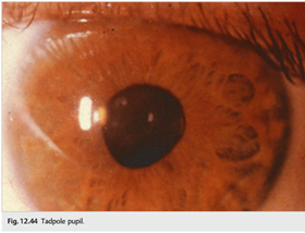
42. What is a tadpole pupil?
Tadpole pupil is an irregular pupil that resembles a tadpole. It is a benign phenomenon that is spontaneously reversible. The pupil undergoes sectoral dilation lasting for a few minutes before returning to normal (segmental spasm of the iris dilator muscle). Tadpole pupil may occur multiple times for several days or a week and then disappear.
43. What are 6 causes of light-near dissociation?
1. Severe loss of afferent light input to both eyes (retina, optic nerves, chiasm),
2. Ocular surgery (laser panretinal photocoagulation, cryotherapy orbital surgery),
3. Adie tonic pupil,
4. Argyll Robertson pupils,
5. Aberrant regeneration of the 3rd nerve,
6. Peripheral neuropathy
44. What the characteristics of the Argyll Robertson pupil?
Argyll Robertson pupils are small (<2mm), irregular pupils (almost always bilateral). They are characterized by no reaction to light, normal near response (light-near dissociation), often with iris atrophy, iris transillumination defects, and poor dilation with mydriatic drops. The condition is classically due to tertiary syphilis, diabetes, and may happen with encephalitis.
45. What is the mechanism in light-near dissociation due to Argyll Robertson pupils?
Most descriptions of the AR pupil do not mention segmental iris sphincter constriction, or slow, sustained constriction with a near vision effort. Such features are considered typical of the light-near dissociation of Adie syndrome and of neuropathic tonic pupils, where damage to the ciliary ganglion or ciliary nerves is believed to be the mechanism. Because the AR pupil lacks these features, it has been attributed to a dorsal midbrain lesion that interrupts the pupillary light reflex pathway but spares the more ventral pupillary near reflex pathway. However, lesions in this region have not been reliably demonstrated in syphilis. Resolving the issue about the location of the syphilitic lesion that produces the AR pupil will depend on careful examination of patients with techniques designed to disclose segmental palsy of the iris. If segmental iris sphincter palsy is found and the light-near dissociation has tonic features, one must conclude that the mechanism of the pupil disorder is a ciliary (peripheral) rather than a midbrain (central) denervation. Until better evidence settles the localization of the AR pupil, it is appropriate to screen patients with bilateral tonic pupils for syphilis.2
46. Where is the lesion in a patient with light-near dissociation due to Adie tonic pupil?
The ciliary ganglion
47. What is the mechanism in light-near dissociation due to aberrant regeneration of the 3rd nerve?
Aberrant reinnervation of the iris sphincter by fibers meant for the extraocular muscles or the ciliary body.
48. What is the mechanism in light-near dissociation due to severe vision loss?
Damage to retina or optic nerve results in decreased light response, but the neurologic pathways for near response are intact.
49. What is the mechanism in light-near dissociation due to laser panretinal photocoagulation or cryotherapy?
Aberrant reinnervation of the iris sphincter following damage to the short posterior ciliary nerves
50. Where is the lesion in a patient with light-near dissociation due to peripheral neuropathy?
There is axonal loss in the short posterior ciliary nerves.
The information below is from Neuro-ophthalmology Illustrated-2nd Edition. Biousse V and Newman NJ. 2012. Thieme
12.5 Other Pupillary Abnormalities
12.5.1 Tadpole Pupil
Tadpole pupil is an irregular pupil that resembles a tadpole (▶Fig. 12.44). It is a benign phenomenon that is spontaneously reversible. The pupil undergoes sectoral dilation lasting for a few minutes before returning to normal (segmental spasm of the iris dilator muscle). Tadpole pupil may occur multiple times for several days or a week and then disappear.
12.5.2 Midbrain Corectopia
Midbrain corectopia refers to eccentric or oval pupils occasionally seen in patients with rostral midbrain lesions.
12.5.3 Argyll Robertson Pupils
Argyll Robertson pupils are small (<2mm), irregular pupils (almost always bilateral). They are characterized by no reaction to light, normal near response (light-near dissociation), often iris atrophy and iris transillumination defects, and poor dilated with drops. The condition is classically described in patients with tertiary syphilis, is common in diabetes, and may happen in encephalitis.
12.5.4 Light-Near Dissociation
Light-near dissociation refers to pupils that do not react to light but react to near stimuli (▶Table 12.7).

The Argyll Robertson pupil.
Thompson HS1, Kardon RH. J Neuroophthalmol. 2006;26(2):134-8
Abstract
The Argyll Robertson (AR) pupil has been defined as a pupil that is small and constricts poorly to direct light but briskly when a target within reading distance is viewed (“light-near dissociation”). Most descriptions of the AR pupil do not mention segmental iris sphincter constriction, or slow, sustained constriction with a near vision effort. Such features are considered typical of the light-near dissociation of Adie syndrome and of neuropathic tonic pupils, where damage to the ciliary ganglion or ciliary nerves is believed to be the mechanism. Because the AR pupil lacks these features, it has been attributed to a dorsal midbrain lesion that interrupts the pupillary light reflex pathway but spares the more ventral pupillary near reflex pathway. However, lesions in this region have not been reliably demonstrated in syphilis. Resolving the issue about the location of the syphilitic lesion that produces the AR pupil will depend on careful examination of patients with techniques designed to disclose segmental palsy of the iris. If segmental iris sphincter palsy is found and the light-near dissociation has tonic features, one must conclude that the mechanism of the pupil disorder is a ciliary (peripheral) rather than a midbrain (central) denervation. Until better evidence settles the localization of the AR pupil, it is appropriate to screen patients with bilateral tonic pupils for syphilis.
References:
1. Neuro-ophthalmology Illustrated-2nd Edition. Biousse V and Newman NJ. 2012. Thieme
2. The Argyll Robertson pupil.Thompson HS1, Kardon RH.J Neuroophthalmol. 2006;26(2):134-8.
These questions are archived at https://neuro-ophthalmology.stanford.edu
Follow https://twitter.com/NeuroOphthQandA to be notified of new neuro-ophthalmology questions of the week.
Please send feedback, questions, and corrections to tcooper@stanford.edu.