Questions:
31. What is akinetopsia?
32. How does one test for akinetopsia?
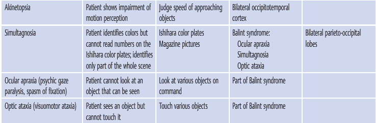
33. Where is the lesion in akinetopsia?
34. What is simultagnosia?
35. How does one test for simultagnosia?
36. What clinical signs are associated with simultagnosia?
37. Where is the lesion in simultagnosia?
____________________________________________________
Questions with answers:
31. What is akinetopsia?
It is an extremely rare Higher Cortical Disorder in which the patient shows impairment of motion perception (motion blindness).
32. How does one test for akinetopsia?
Have the patient judge the speed of approaching objects. The patient will have difficulty in the perception of objects in motion, despite being able to see stationary objects without issue.
33. Where is the lesion in akinetopsia?
Bilateral occipitotemporal cortex
34. What is simultagnosia?
It is the Higher Cortical Disorder in which the patient inability to grasp the entire meaning of a picture despite an intact capacity to recognize the picture’s individual constituent elements.
35. How does one test for simultagnosia?
Present magazine pictures to the patient and ask the patient to describe the picture. If the patient is presented with a picture of a table containing both food and various utensils, a patient will report seeing only one item, such as a spoon. If the patient’s attention is redirected to another object in the scene, such as a glass, the patient will report that they see the glass but no longer see the spoon.
36. What clinical signs are associated with simultagnosia?
Part of Balint syndrome: Ocular apraxia, Simultagnosia, Optic ataxia
37. Where is the lesion in simultagnosia?
Bilateral parieto-occipital lobes
The information below is from Neuro-ophthalmology Illustrated-2nd Edition. Biousse V and Newman NJ. 2012. Thieme
10 Disorders of Higher Cortical Function
Unlike the anterior visual and geniculocalcarine pathways that deliver basic visual information from the eyes to the occipital cortex, association cortical visual areas (higher cortical areas) perform the more complex interpretation of visual information. Many of the syndromes of higher cortical dysfunction are secondary to a disconnection of the flow of visual information between the striate cortex and other cortical regions. When these areas are damaged, visual processing is abnormal despite often normal visual acuity and visual fields.
This chapter focuses on some of the main visual disorders of higher cortical function, particularly their clinical and radiologic findings and causes, commonly encountered in neuro-ophthalmology.
10.1 Classification
Disorders of higher cortical function are often grouped into two processing streams. The first stream, the inferior (ventral) or occipitotemporal pathway for object recognition, extends from below the calcarine fissure into the adjacent temporal lobe. It facilitates object recognition and color perception. Disorders here include achromatopsia, prosopagnosia, alexia, and topographagnosia.
The second stream, the superior (dorsal) or occipitoparietal pathway for object localization extends from the upper bank of the calcarine fissure into the adjacent parietal lobe. It processes visuospatial attributes, including location and motion. Disorders here include akinetopsia, Balint syndrome (simultagnosia, ocular apraxia, and optic ataxia), and hemineglect.
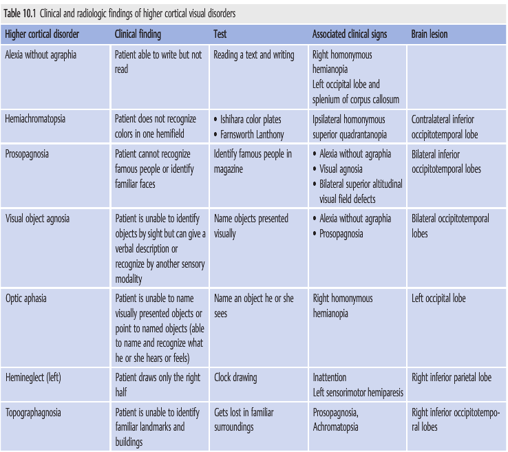
▶Table 10.1 lists the clinical findings, recommended tests, associated clinical signs, and lesions associated with these higher cortical disorders.
Reference: 1. Neuro-ophthalmology Illustrated-2nd Edition. Biousse V and Newman NJ. 2012. Thieme
These questions are archived at https://neuro-ophthalmology.stanford.edu
Follow https://twitter.com/NeuroOphthQandA to be notified of new neuro-ophthalmology questions of the week.
Please send feedback, questions, and corrections to tcooper@stanford.edu.