Questions:
15. What is visual object agnosia?
16. How does one test for visual object agnosia?
17. What clinical signs are associated with object agnosia?
18. Where is the lesion in visual object agnosia?
19. What is optic aphasia?
20. How does one test for optic aphasia?
21. What clinical signs are associated with optic aphasia?
22. Where is the lesion in optic aphasia?
____________________________________________________
Questions with answers:
15. What is visual object agnosia?
It is the Higher Cortical Disorder in which the patient is unable to identify objects by sight but can give a verbal description or recognize the object by another sensory modality.
16. How does one test for visual object agnosia?
Have the patient attempt to name objects presented visually.
17. What clinical signs are associated with object agnosia?
Alexia without agraphia and prosopagnosia
18. Where is the lesion in visual object agnosia?
Bilateral occipitotemporal lobes
19. What is optic aphasia?
It is the Higher Cortical Disorder in which the patient is unable to name visually presented objects or point to named objects. These patients can name and recognize what he or she hears or feels.
20. How does one test for optic aphasia?
Have the patient attempt to name an object he or she sees.
21. What clinical signs are associated with optic aphasia?
Right homonymous hemianopia
22. Where is the lesion in optic aphasia?
Left occipital lobe
The information below is from Neuro-ophthalmology Illustrated-2nd Edition. Biousse V and Newman NJ. 2012. Thieme
10 Disorders of Higher Cortical Function
Unlike the anterior visual and geniculocalcarine pathways that deliver basic visual information from the eyes to the occipital cortex, association cortical visual areas (higher cortical areas) perform the more complex interpretation of visual information. Many of the syndromes of higher cortical dysfunction are secondary to a disconnection of the flow of visual information between the striate cortex and other cortical regions. When these areas are damaged, visual processing is abnormal despite often normal visual acuity and visual fields.
This chapter focuses on some of the main visual disorders of higher cortical function, particularly their clinical and radiologic findings and causes, commonly encountered in neuro-ophthalmology.
10.1 Classification
Disorders of higher cortical function are often grouped into two processing streams. The first stream, the inferior (ventral) or occipitotemporal pathway for object recognition, extends from below the calcarine fissure into the adjacent temporal lobe. It facilitates object recognition and color perception. Disorders here include achromatopsia, prosopagnosia, alexia, and topographagnosia.
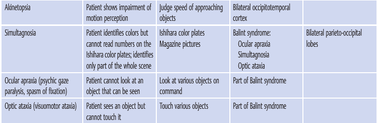
The second stream, the superior (dorsal) or occipitoparietal pathway for object localization extends from the upper bank of the calcarine fissure into the adjacent parietal lobe. It processes visuospatial attributes, including location and motion. Disorders here include akinetopsia, Balint syndrome (simultagnosia, ocular apraxia, and optic ataxia), and hemineglect.
▶Table 10.1 lists the clinical findings, recommended tests, associated clinical signs, and lesions associated with these higher cortical disorders.

Reference: 1. Neuro-ophthalmology Illustrated-2nd Edition. Biousse V and Newman NJ. 2012. Thieme
These questions are archived at https://neuro-ophthalmology.stanford.edu
Follow https://twitter.com/NeuroOphthQandA to be notified of new neuro-ophthalmology questions of the week.
Please send feedback, questions, and corrections to tcooper@stanford.edu.