Questions:
17. Once optic disc edema is confirmed, what should be determined?
18. In anterior optic neuropathy with disk edema, is visual acuity usually decreased?
19. In papilledema is visual acuity usually decreased?
20. In anterior optic neuropathy with disk edema is color vision usually decreased?
21. In papilledema is color vision usually decreased?
22. What are the usual characteristics of visual field defects in anterior optic neuropathy with disk edema?
23. What are the usual characteristics of visual field defects in papilledema
24. Is anterior optic neuropathy with disk edema usually unilateral?
25. Is papilledema usually unilateral?
26. What findings are often associated with papilledema?
27. Does the absence of disc edema rule-out raised intracranial pressure in a patient presenting with headache?
28. What visual symptoms are associated with papilledema?
29. When is central visual acuity loss experienced in papilledema, early or late?
30. What type of visual field defect is found initially with papilledema?
31. What type of visual field loss is experienced in long-standing papilledema?
32. Can visual loss from papilledema occur with any cause of papilledema?
33. What should be assessed next in a patient found to have papilledema?
_____________________________________________
Questions with answers:
17. Once optic disc edema is confirmed, what should be determined?
Once optic disc edema is confirmed, it should be determined whether it is related to an optic nerve disorder (optic neuropathy) or to raised intracranial pressure (papilledema).
18. In anterior optic neuropathy with disk edema. is visual acuity usually decreased?
Yes
19. In papilledema is visual acuity usually decreased?
No
20. In anterior optic neuropathy with disk edema is color vision usually decreased?
Yes
21. In papilledema is color vision usually decreased?
No
22. What are the usual characteristics of visual field defects in anterior optic neuropathy with disk edema?
Central, arcuate, and altitudinal defects
23. What are the usual characteristics of visual field defects in papilledema? Enlarged blind spots, nasal defects, and general constriction
24. Is anterior optic neuropathy with disk edema usually unilateral?
Yes
25. Is papilledema usually unilateral?
No
26. What findings are often associated with papilledema?
Symptoms of increased ICP (headache, nausea, diplopia from 6th nerve palsies, pulsatile tinnitus, transient visual obscurations) and focal neurologic signs or symptoms may be present.
27. Does the absence of disc edema rule-out raised intracranial pressure in a patient presenting with headache?
Although papilledema is a reliable sign of raised intracranial pressure, the absence of disc edema does not rule out raised intracranial pressure in a patient presenting with headache.
28. What visual symptoms are associated with papilledema?
Patients may complain of “flashing lights” or transient visual obscurations (brief episodes of visual loss occurring in one or both eyes). These are often precipitated by changes in posture, such as standing up after bending over.
29. When is central visual acuity loss experienced in papilledema, early or late?
In papilledema central visual acuity is normal until late.
30. What type of visual field defect is found initially with papilledema?
Blind spot enlargement and nasal defects are common initially.
31. What type of visual field loss is experienced in long-standing papilledema?
Patients develop insidious progressive visual field constriction.
32. Can visual loss from papilledema occur with any cause of papilledema
Visual loss from papilledema can occur with any cause of papilledema. Hence it is very important to look for papilledema in all patients with headache or known hydrocephalus, brain tumor, or meningitis and to provide urgent treatment to prevent visual loss when papilledema is present.
33. What should be assessed next in a patient found to have papilledema?
Blood pressure measurement to rule-out malignant hypertension followed by emergent neuro-imaging (preferably brain MRI & MRV with contrast).
The information below is from Neuro-ophthalmology Illustrated-2nd Edition. Biousse V and Newman NJ. 2012. Thieme
9.4 Evaluation of the Patient with Disc Edema
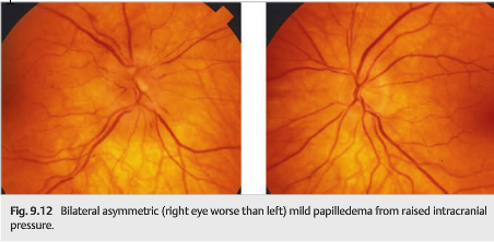
Once optic disc edema is confirmed, it should be determined whether it is related to an optic nerve disorder (optic neuropathy) or to raised intracranial pressure. Papilledema Is the term used to describe optic disc edema resulting from raised intracranial pressure (▶Fig. 9.12). All other optic disc edema is termed disc edema or swollen optic nerve. ▶Table 9.2 compares the characteristics of disc edema from anterior optic neuropathy with those from raised intracranial pressure.

Most disorders producing raised intracranial pressure are life-threatening emergencies. The finding of papilledema should prompt an immediate workup, ideally in a specialized center with up-to-date neuroimaging, as well as neurologic and ophthalmological consultations.
The workup should include the following:
● Looking for an underlying neurologic process
● Careful evaluation of the visual function (visual acuity and formal visual field testing), as papilledema can result in permanent visual loss from secondary optic atrophy
● Checking blood pressure (severe systemic hypertension or malignant hypertension may produce bilateral disc edema that mimics papilledema)
Pearls
Although papilledema is a reliable sign of raised intracranial pressure, the absence of disc edema does not rule out raised intracranial pressure in a patient presenting with headache.
The mechanisms responsible for raised intracranial pressure and papilledema are as follows:
● Hydrocephalus (▶Fig. 9.13)

● Intracranial mass
○ Tumor, abscess ((▶Fig. 9.14)

○ Intracerebral hemorrhage
○ Subdural/epidural hemorrhage
○ Large vascular malformation
● Meningeal process
○ IInfectious
○ Inflammatory
○ Neoplastic
● Increased venous pressure
● Cerebral venous thrombosis
● Idiopathic intracranial hypertension
Increased venous pressure produces symptoms and signs of raised intracranial pressure, including papilledema.
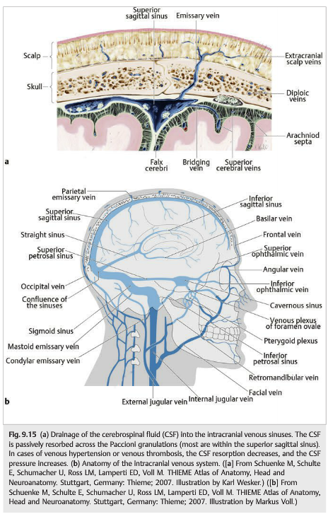
The causes of increased venous pressure include all of the causes of decreased venous return (▶Fig. 9.15):
● Right cardiac insufficiency
● Pulmonary hypertension
● Sleep apnea syndrome
● Superior vena cava syndrome
● Jugular vein occlusion
● Dural fistula
● Cerebral venous stenosis
● Cerebral venous thrombosis
Cerebral venous thrombosis is a classic cause of raised intracranial pressure (▶Fig. 9.16; see also Chapter 20). Patients may present with isolated raised intracranial pressure, thereby mimicking idiopathic intracranial hypertension. Early recognition may prevent a devastating stroke and visual loss from chronic papilledema.

When evaluating a patient with presumed papilledema (raised intracranial pressure), neuroimaging needs to be obtained emergently to rule out an intracranial process (▶Fig. 9.17). Magnetic resonance imaging (MRI) with contrast of the brain is the ideal test and is the most sensitive to detect intracranial masses, infiltrative and meningeal processes, and cerebral venous thrombosis. Computed tomography (CT )without contrast, which is often the test of choice in the emergency room, is in most cases not helpful in these patients, unless it is followed by brain MRI. Indeed, the CT is helpful to detect intracranial hemorrhages, hydrocephalus, and large mass lesions, but it does not rule out any of the other intracranial lesions. Patients with a normal head CT should be investigated further with brain MRI (see Chapter 4). A normal brain MRI scan in the setting of papilledema suggests a meningeal process, venous hypertension, or idiopathic intracranial hypertension as the cause of raised intracranial pressure. A Lumbar puncture with measurement of the cerebrospinal fluid (CSF) opening pressure and CSF analysis should always be performed.

9.5 Classification and Progression of Papilledema
Patients with papilledema often have no visual symptoms initially. They may complain of “flashing lights” or transient visual obscurations (brief episodes of visual loss occurring in one or both eyes), often precipitated by changes in posture, such as standing up after bending over. Untreated chronic papilledema results in visual loss: central visual acuity is normal until late, and patients develop insidious progressive visual field constriction (▶Fig. 9.18,▶Fig. 9.19,▶Fig. 9.20,▶Fig. 9.21,▶Fig. 9.22).


Formal visual field testing (Humphrey perimetry shown in ▶Fig. 9.23) is often abnormal in papilledema. Blind spot enlargement and nasal defects are common initially (top, ▶Fig. 9.23). They may progress, usually circumferentially, to involve the central 30 degrees of the visual field (middle). Severe devastating visual field loss (bottom) is often permanent if raised intracranial pressure is not promptly treated (note that even with the severe visual field loss seen in the bottom example, visual acuity was still relatively preserved at 20/25 OD [right eye] and 20/40 OS [left eye]).

Pearls
Visual loss from papilledema happens with any cause of papilledema. Hence it is very important to look for papilledema in all patients with headache or known hydrocephalus, brain tumor, or meningitis and to prevent visual loss when papilledema is present.
Reference: 1. Neuro-ophthalmology Illustrated-2nd Edition. Biousse V and Newman NJ. 2012. Thieme
These questions are archived at https://neuro-ophthalmology.stanford.edu
Follow https://twitter.com/NeuroOphthQandA to be notified of new neuro-ophthalmology questions of the week.
Please send feedback, questions, and corrections to tcooper@stanford.edu.