Questions:
130. What type of traumatic optic neuropathy may require emergent treatment?
131. What is the management of indirect traumatic optic neuropathy?
132. What should be done in all cases of head trauma?
133. What potential alternative causes of optic neuropathy should be ruled out when considering the diagnosis of low-tension glaucoma?
134. Are optic nerve head drusen usually unilateral or bilateral?
135. Do patients with optic nerve head drusen experience transient visual obscurations?
136. What are optic nerve head drusen?
137. What should be done if a patient presents with visual symptoms of optic neuropathy and is found to have optic nerve head drusen?
138. What is the most common congenital optic nerve anomaly?
139. What are the characteristics of optic nerve hypoplasia?
140. What conditions are associated with optic nerve hypoplasia?
141. What is an optic disk coloboma?
142. What conditions are associated with optic disk coloboma?
143. What are the characteristics of the “morning glory” disk anomaly?
144. What is the mechanism behind the morning glory disk anomaly?
145. What conditions are associated with the “morning glory” disk anomaly?
146. What is the mechanism of optic pit development?
147. What are the characteristics of an optic pit?
148. What are the characteristics of the tilted disk anomaly?
149. What is the usual field defect seen with myelinated retinal nerve fibers around the disk?
____________________________________________________
Questions with answers:
130. What type of traumatic optic neuropathy may require emergent treatment?
Direct traumatic optic neuropathies may require emergent surgical treatment to decompress the optic nerve and treat the fractures.
131. What is the management of indirect traumatic optic neuropathy?
The management of indirect traumatic optic neuropathy is controversial. Although visual loss may be devastating and permanent, vision may also recover spontaneously. There is no indication for surgical decompression of the injured optic nerve, and corticosteroids (even very high doses) are not helpful and may even be harmful when administered more than 8 hours after injury. In addition, the use of corticosteroids should be avoided in patients with systemic traumatic injuries and traumatic brain injury.
132. What should be done in all cases of head trauma?
The visual acuity and the pupils should be evaluated in all head trauma patients. If abnormal, obtain a CT of the brain and orbits without contrast and with bone windows.
133. What potential alternative causes of optic neuropathy should be ruled out when considering the diagnosis of low-tension glaucoma?
Low-tension glaucoma is a diagnosis of exclusion as other causes of optic neuropathies particularly compressive need to be ruled out by neuroimaging. Dominant (or Kjer) optic atrophy is often misdiagnosed as low-tension glaucoma.
134. Are optic nerve head drusen usually unilateral or bilateral?
Bilateral
135. Do patients with optic nerve head drusen experience transient visual obscurations?
Some patients with optic nerve head drusen have brief episodes of transient visual obscurations.
136. What are optic nerve head drusen?
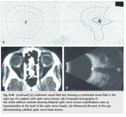
Drusen are small, calcific concretions present in the optic nerve of 1 to 2% of the population. Autosomal dominant transmission is suspected in most cases. Optic nerve head drusen are usually bilateral and they may be asymmetric. They may be visible on fundus examination in teenagers and in adults (often buried in younger children) and may slowly increase over time. They are often asymptomatic and discovered during routine fundus examination.
137. What should be done if a patient presents with visual symptoms of optic neuropathy and is found to have optic nerve head drusen?
Incidentally found optic nerve drusen are common and are usually asymptomatic. Symptomatic patients should undergo the usual evaluation of optic neuropathy. In some of these cases, optic nerve drusen are a “red herring”.
138. What is the most common congenital optic nerve anomaly?
Optic nerve hypoplasia
139. What are the characteristics of optic nerve hypoplasia?
It is the most common congenital optic nerve anomaly. It is characterized by a small optic nerve (reduced diameter), sometimes with a peripapillary halo (double ring sign), and may be unilateral or bilateral.
140. What conditions are associated with optic nerve hypoplasia?
1. Midline developmental brain abnormalities (e. g. Septo-optic dysplasia (de Morsier syndrome): absent septum pellucidum, thin corpus callosum, hypopituitarism with pituitary ectopia)
2. Numerous congenital syndromes including albinism, aniridia and Duane syndrome
3. Maternal diabetes
4. Fetal alcohol syndrome
5. Drugs or substances taken by the mother during pregnancy (phenytoin, quinine, phencyclidine hydrochloride, lysergic acid diethylamide, and alcohol)
141. What is an optic disk coloboma?
Optic disk coloboma is faulty closure of the embryonic fetal fissure of the optic stalk and cup.
142. What conditions are associated with optic disk coloboma?
It may be isolated or associated with coloboma of the iris, retina, and choroid. A rare association with forebrain abnormalities (basal encephalocele) warrants routine neuroimaging of patients with disk colobomas.
143. What are the characteristics of the “morning glory” disk anomaly?
They include congenital funnel-shaped excavation of the posterior pole of the fundus (the optic disk lies within the excavation), severe peripapillary pigmentary changes, and anomalous straightening of the retinal vasculature.
144. What is the mechanism behind the morning glory disk anomaly?
The mechanism behind “morning glory” disk anomaly is debated, but it results at least partially from faulty closure of the embryonic fetal fissure of the optic stalk and cup.
145. What conditions are associated with the “morning glory” disk anomaly?
A basal encephalocele often containing the optic chiasm and hypothalamus, protrudes through a defect in the sphenoid bone. Children with this structural defect often have dysmorphic features, including a wide head, flat nose, hypertelorism and a midline notch in the upper lip.
Many patients with transsphenoidal encephalocele also have callosal agenesis, leading to dilation of the lateral ventricles. It is also associated with cerebrovascular anomalies, including hypoplasia of the cerebral arteries, Moyamoya, PHACE syndrome (posterior fossa malformation, large facial hemangioma, arterial anomalies, cardiac anomalies, eye anomalies) and Neurofibromatosis type 2.
146. What is the mechanism of optic pit development?
Optic pit results from faulty closure of the embryonic fetal fissure of the optic stalk and cup.
147. What are the characteristics of an optic pit?
It is characterized by a small excavation of the neuroretinal rim of the optic disk and usually involves the inferotemporal portion of the optic nerve. The missing area often is associated with an arcuate visual field defect. Optic pits may be associated with serous detachment of the macula causing decreased visual acuity.
148. What are the characteristics of the tilted disk anomaly?
The tilted disk anomaly arises when the optic nerve enters the sclera at an oblique angle. It is common in high myopes and often results in relative bitemporal visual field defects that do not respect the vertical meridian.
149. What is the usual field defect seen with myelinated retinal nerve fibers around the disk?
Intraocular myelination of retinal nerve fibers is seen in < 1% of the population. Visual field defects are seen only with extensive intraocular myelination, usually an enlarged blind spot when the myelination surrounds the optic disk.
The information below is from Neuro-ophthalmology Illustrated-2nd Edition. Biousse V and Newman NJ. 2012. Thieme
8.9 Traumatic Optic Neuropathy
Traumatic optic neuropathy is an uncommon but potentially devastating complication of head injury. It should always be suspected in any patient with evidence of optic nerve dysfunction (e.g., otherwise unexplained decreased visual acuity, RAPD, or dyschromatopsia) following head trauma. Because of associated neurologic deficits and other traumatic injuries, the diagnosis is often delayed. However, systematic examination of the pupils in the emergency room (looking for a RAPD) should allow early diagnosis, even in unresponsive patients.
8.9.1 Mechanism
The optic nerve may be injured directly by an orbital foreign body or by a bone fragment in case of orbital fracture (direct traumatic optic neuropathy; ▶Fig. 8.41), or indirectly, as a result of concussive forces to the head, particularly the forehead (indirect traumatic optic neuropathy; (▶Fig. 8.42). The latter causes both a mechanical and ischemic insult to the optic nerve, likely at the level of the optic canal.


8.9.2 Treatment
Direct traumatic optic neuropathies usually require emergent surgical treatment to decompress the optic nerve and treat the fracture.
The management of indirect traumatic optic neuropathy is controversial. Although Visual loss may be devastating and permanent, vision may also recover spontaneously. There is no indication for surgical decompression of the injured optic nerve, and corticosteroids (even very high doses) are not helpful and may even be harmful when administered more than 8 hours after injury. In addition, the use of corticosteroids should be avoided in patients with systemic traumatic injuries and traumatic brain injury who are at risk for infectious complications.
Pearls
Check the visual acuity and the pupils of all trauma patients. If abnormal, request an ophthalmology consultation. Obtain a CT scan of the brain and orbits without contrast and with bone windows.
8.11 Optic Nerve Anomalies
8.11.1 Optic Nerve Head Drusen
Drusen are small, calcific concretions present in the optic nerve of 1 to 2% of the population (▶Fig. 8.44). Autosomal dominant transmission is suspected in most cases. Optic nerve head drusen are usually bilateral, but they may be asymmetric. They may be visible on fundus examination in teenagers and in adults (often buried in younger children) and may slowly increase over time. They are often asymptomatic and discovered during routine fundus examination. Some patients have brief episodes of transient visual obscurations. The drusen may mimic disc edema when they are buried in the optic nerve head. They may result in peripheral visual field defects, which may worsen slowly over time (optic nerve head drusen usually do not produce central visual loss). The diagnosis is easy when the drusen are superficial and seen on funduscopic examination. Buried drusen can be seen with autofluorescence imaging of the optic nerves, OCT, B-scan ultrasonography and CT scan of the orbits showing calcifications in the optic nerve heads (▶Fig. 8.44).

Pearls
Incidentally found optic nerve drusen are common and are usually asymptomatic. Symptomatic patients should undergo an evaluation for other causes of optic neuropathies because, in some cases, the optic nerve drusen are a red herring.
8.11.2 Congenital Disc Anomalies
Congenital disc anomalies may be isolated or associated with systemic disorders or malformations. The level of visual loss associated with congenital disc anomalies varies from minimal visual dysfunction to total blindness. In childhood, the most common presentation of unilateral disc anomaly is strabismus, whereas those with bilateral disc anomalies more frequently present with poor vision or nystagmus. Some may also be diagnosed during adulthood on a routine funduscopic examination.
Optic Nerve Hypoplasia
Optic nerve hypoplasia (▶Fig. 8.45) is the most common congenital optic nerve anomaly. It is characterized by a small optic nerve (reduced diameter), sometimes with peripapillary halo (double ring sign), and may be unilateral or bilateral.

Pearl
A brain MRI scan looking for septo-optic dysplasia and pituitary ectopia should be obtained in all cases of optic nerve hypoplasia. All patients with septo-optic dysplasia or pituitary ectopia need an endocrine evaluation looking for panhypopituitarism, which may be life threatening when not diagnosed.
A brain MRI scan looking for septo-optic dysplasia and pituitary ectopia should be obtained in all cases of optic nerve hypoplasia. All patients with septo-optic dysplasia or pituitary ectopia need an endocrine evaluation looking for panhypopituitarism, which may be life threatening when not diagnosed.
Classic systemic and teratogenic associations with optic nerve hypoplasia include the following:
● Midline developmental abnormalities
○ Septo-optic dysplasia (de Morsier syndrome): absent septum pellucidum, thin corpus callosum, hypopituitarism with pituitary ectopia
● Albinism, aniridia, Duane syndrome, and numerous other congenital ocular syndromes
● Maternal diabetes
● Fetal alcohol syndrome
● Drugs or substances taken by the mother during pregnancy, such as phenytoin, quinine, phencyclidine hydrochloride (PCP), lysergic acid diethylamide (LSD), and alcohol
Optic Disc Coloboma
Optic disc coloboma is faulty closure of the embryonic fetal fissure of the optic stalk and cup. It is isolated or associated with coloboma of the iris, retina, and choroid. A rare association with forebrain abnormalities (basal encephalocele) warrants routine neuroimaging of patients with colobomas. Characteristic features of the optic disc coloboma(▶Fig. 8.46) include excavation within the optic disc, asymmetric defect, vision varying based on size and location of the coloboma within the disc, and minimal peripapillary pigmentary changes. Retinal vasculature is normal.

Morning Glory Disc Anomaly
The mechanism behind “morning glory” disc anomaly is debated, but it results at least partially from faulty closure of the embryonic fetal fissure of the optic stalk and cup. Its association with transsphenoidal basal encephaloceles warrants routine neuroimaging of patients with such anomalies. Characteristic features of morning glory disc anomaly (▶Fig. 8.47) include congenital funnel-shaped excavation of the posterior pole of the fundus (the optic disc lies within the excavation), severe peripapillary pigmentary changes, and anomalous straightening of the retinal vasculature.

Optic Pit
Optic pit (▶Fig. 8.48) results from faulty closure of the embryonic fetal fissure of the optic stalk and cup (same as coloboma, which is a more severe variant). It is characterized by a small excavation of the neuroretinal rim of the optic disc and usually involves the inferotemporal portion of the optic nerve. The missing area is often associated with an arcuate visual field defect. Optic pit may be associated with serous detachment of the macula, causing decreased visual acuity.

Tilted Disc Anomaly
Tilted disc anomaly (▶Fig. 8.49) arises when the optic nerve enters the sclera at an oblique angle. It is common in high myopes and often results in relative bitemporal visual field defects that do not respect the vertical meridian.

Myelinated Nerve Fibers
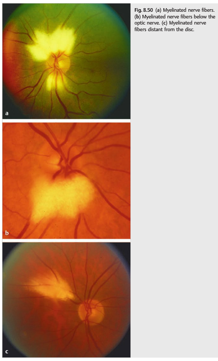
The optic nerve fibers behind the lamina cribrosa are normally myelinated. Usually, myelin does not enter the eye. Intraocular myelination of retinal nerve fibers is seen in <1% of the population (▶Fig. 8.50). Visual field defects are seen only with extensive intraocular myelination, usually in the form of an enlarged blind spot when the myelination surrounds the optic disc.
Reference: 1. Neuro-ophthalmology Illustrated-2nd Edition. Biousse V and Newman NJ. 2012. Thieme
These questions are archived at https://neuro-ophthalmology.stanford.edu
Follow https://twitter.com/NeuroOphthQandA to be notified of new neuro-ophthalmology questions of the week.
Please send feedback, questions, and corrections to tcooper@stanford.edu.