Questions:
101. What are the characteristics of hereditary optic neuropathies?
102. When should hereditary optic neuropathies be suspected?
103. What is the most common hereditary optic neuropathy?
104. What misdiagnosis is often made in dominant optic atrophy?
105. What is the typical disk appearance in dominant optic atrophy?
106. Can hereditary optic atrophies have other neurologic or systemic signs?
107. Are hereditary optic atrophies associated with degenerative or developmental diseases?
108. How is Leber hereditary optic neuropathy (LHON) inherited?
109. What are the clinical findings in Leber hereditary optic neuropathy (LHON)?
110. What can be associated with Leber hereditary optic neuropathy (LHON?
111. Is there a genetic blood test for Leber hereditary optic neuropathy (LHON)?
112. Does spontaneous vision improvement occur in some patients with Leber hereditary optic neuropathy (LHON)?
113. What test should be done on patients confirmed as having Leber hereditary optic neuropathy (LHON)?
114. Which hereditary optic atrophy often has disk cupping in addition to bilateral temporal disk pallor?
115. What are the characteristics of Autosomal Dominant Optic Atrophy (DOA)?
____________________________________________________
Questions with answers:
101. What are the characteristics of hereditary optic neuropathies? Hereditary optic neuropathies usually produce bilateral painless optic neuropathies and permanent visual loss.
102. When should hereditary optic neuropathies be suspected?
They should be suspected in any case of optic neuropathy of unknown cause, even in patients without a family history of visual loss.
103. What is the most common hereditary optic neuropathy?
The most common cause of hereditary optic neuropathy is dominant (Kjer) optic atrophy.
104. What misdiagnosis is often made in dominant (Kjer) optic atrophy?
It is often misdiagnosed as glaucoma in adults.
105. What is the typical disk appearance in dominant (Kjer) optic atrophy? Bilateral temporal disk pallor and disks often appear cupped.
106. Can hereditary optic atrophies have other neurologic or systemic signs?
Yes. Hereditary optic atrophies can have other neurologic of systemic signs. They can also be a manifestation of hereditary degenerative or developmental diseases.
Hereditary Optic Atrophy with Other Neurologic or Systemic Signs
The main types of hereditary optic atrophy with other neurologic or systemic signs include the following:
● Wolfram/diabetes insipidus, diabetes mellitus, optic atrophy, and deafness (DIDMOAD) syndrome
● Autosomal dominant optic atrophy and deafness
● Autosomal dominant optic atrophy with associated deafness and other neurologic signs
● Behr syndrome
107. Are hereditary optic atrophies associated with degenerative or developmental diseases? Yes, hereditary optic atrophies can be associated with degenerative or developmental diseases.
Optic Neuropathy as a Manifestation of Hereditary Degenerative or Developmental Diseases
Hereditary degenerative or developmental diseases associated with optic neuropathy include the following:
● Hereditary ataxias
○ Friedreich ataxia
○ Spinocerebellar ataxias
● Hereditary polyneuropathies
○ Charcot–Marie–Tooth disease
○ Familial dysautonomia (Riley–Day syndrome)
● Hereditary spastic paraplegias
● Hereditary muscular dystrophies
● Storage diseases and cerebral degenerations of childhood
● Mitochondrial diseases of childhood
○ Leigh syndrome
○ Mitochondrial myopathy, encephalopathy, lactic acidosis, and stroke-like episodes(MELAS) syndrome
○ Myoclonic epilepsy and ragged red fibers (MERRF) syndrome
○ Chronic progressive external ophthalmoplegia (CPEO)/Kearns–Sayre syndrome
108. How is Leber hereditary optic neuropathy (LHON) inherited?
It is a maternally inherited mitochondrial disease.
109. What are the clinical findings in Leber hereditary optic neuropathy (LHON)?
LHON is a bilateral sequential or simultaneous optic neuropathy that occurs predominantly in otherwise healthy young men but may affect either gender at any age. Visual acuity typically deteriorates permanently to levels of 20/200 or worse, and visual fields show central or cecocentral defects. During the acute phase of visual loss, the funduscopic appearance may be normal, or there may be hyperemia and apparent swelling of the optic disk with dilation and tortuosity of the retinal vasculature. Ultimately, the patient will develop optic nerve pallor.
110. What can be associated with Leber hereditary optic neuropathy (LHON?
In LHON, visual loss is most often isolated. It may be associated with cardiac conduction abnormalities, minor neurologic abnormalities, and can be clinically indistinguishable from multiple sclerosis.
111. Is there a genetic blood test for Leber hereditary optic neuropathy (LHON)?
Yes, diagnosis is confirmed by genetic analysis (blood test): screening for three primary mutations in the mitochondrial DNA (positions 11778, 14484, and 3460). Not all subjects with the mutation will develop visual loss (about 20–60% of men and about 4–32% of women). The 11778 mutation is the most common.
112. Does spontaneous vision improvement occur in some patients with Leber hereditary optic neuropathy (LHON)?
Yes, some spontaneous recovery of vision may occur in up to 60% of LHON patients with the 14484 mutation, but only 4% of patients with the 11778 mutation.
113. What test should be done on patients confirmed as having Leber hereditary optic neuropathy (LHON)?
An electrocardiogram looking for cardiac conduction abnormalities.
114. Which hereditary optic atrophy often has disk cupping in addition to bilateral temporal disk pallor?
Autosomal dominant optic atrophy (DOA), or Kjer disease,
115. What are the characteristics of Autosomal dominant optic atrophy (DOA)?
Autosomal dominant optic atrophy (DOA), or Kjer disease, affects both men and women, and 50% of offspring have the mutation. It is characterized by the symmetrical, insidious onset of visual loss in the first decade of life. Visual acuity loss ranges from subtle to 20/200 The diagnosis is usually delayed (patients lose approximately one line of vision per decade on the Snellen visual acuity chart). Color vision is abnormal. Visual fields show cecocentral scotomas. There is bilateral temporal pallor of both optic nerves, which often appear cupped. Other neurologic abnormalities are uncommon, but there may be hearing loss in some families.
The information below is from Neuro-ophthalmology Illustrated-2nd Edition. Biousse V and Newman NJ. 2012. Thieme
8.7 Hereditary Optic Neuropathies
Hereditary optic neuropathies usually produce bilateral painless optic neuropathies and permanent visual loss. They should be suspected in any case of optic neuropathy of unknown cause, even in patients without a family history of visual loss. The most common cause of hereditary optic neuropathy is dominant optic atrophy, which is often overlooked and misdiagnosed as glaucoma in adults. Hereditary Optic neuropathies may present at any age, although they are most common in children and young adults. The optic neuropathy may occur in isolation or may be part of a systemic metabolic or neurologic degenerative disorder. There is no treatment for these optic neuropathies.
8.7.1 Classification of Hereditary Optic Neuropathies
Monosymptomatic Hereditary Optic Neuropathies
There are two types of monosymptomatic hereditary optic neuropathies:
● Leber hereditary optic neuropathy (LHON)
● Dominant (or Kjer) optic atrophy
Hereditary Optic Atrophy with Other Neurologic or Systemic Signs
The main types of hereditary optic atrophy with other neurologic or systemic signs include the following:
● Wolfram/diabetes insipidus, diabetes mellitus, optic atrophy, and deafness(DIDMOAD) syndrome
● Autosomal dominant optic atrophy and deafness
● Autosomal dominant optic atrophy with associated deafness and other neurologic signs
● Behr syndrome
Optic Neuropathy as a Manifestation of Hereditary Degenerative or Developmental Diseases
Hereditary degenerative or developmental diseases associated with optic neuropathy include the following:
● Hereditary ataxias
○ Friedreich ataxia
○ Spinocerebellar ataxias
● Hereditary polyneuropathies
○ Charcot–Marie–Tooth disease
○ Familial dysautonomia (Riley–Day syndrome)
● Hereditary spastic paraplegias
● Hereditary muscular dystrophies
● Storage diseases and cerebral degenerations of childhood
● Mitochondrial diseases of childhood
○ Leigh syndrome
○ Mitochondrial myopathy, encephalopathy, lactic acidosis, and stroke-like episodes(MELAS) syndrome
○ Myoclonic epilepsy and ragged red fibers (MERRF) syndrome
○ Chronic progressive external ophthalmoplegia (CPEO)/Kearns–Sayre syndrome

8.7.2 Leber Hereditary Optic Neuropathy
LHON (▶Fig. 8.36) is a maternally inherited mitochondrial disease due to mitochondrial DNA point mutations; that is, the mutation is transmitted by the mother. All children inherit the mutation, but only women (even if unaffected phenotypically) pass it on to the next generation. Men cannot transmit the disease to their children.
LHON is a bilateral sequential or simultaneous optic neuropathy that occurs predominantly in otherwise healthy young men, but may affect any gender at any age. Visual acuity typically deteriorates permanently to levels of 20/200 or worse, and visual fields show central or cecocentral defects.
During the acute phase of visual loss, the funduscopic appearance may be normal, or there may be hyperemia and apparent swelling of the optic disc with dilation and tortuosity of the retinal vasculature. Ultimately, the patient will develop optic nerve pallor.
In LHON, the visual loss is most often isolated. It may be associated with cardiac conduction abnormalities, minor neurologic abnormalities, or disease clinically indistinguishable from multiple sclerosis.
Diagnosis is confirmed by genetic analysis (blood test): screening for three primary mutations in the mitochondrial DNA (positions 11778, 14484, and 3460). Not all subjects with the mutation will develop visual loss (about 20–50% of men and about4–32% of women). The 11778 mutation is the most common.
Some spontaneous recovery of vision may occur in up to 70% of patients with LHON and the 14484 mutation, but in only 4 to 20% of patients with the 11778 mutation.
Pearls
Patients diagnosed with a mitochondrial disorder such as Leber hereditary optic neuropathy should be screened with electrocardiography looking for cardiac conduction abnormalities.
8.7.3 Autosomal Dominant Optic Atrophy (Kjer Disease)
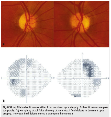
Autosomal dominant optic atrophy (DOA), or Kjer disease, affects both men and women, and 50% of offspring have the mutation (▶Fig. 8.37).
It is characterized by symmetrical, insidious onset of visual loss in the first decade of life. Visual acuity loss is often moderate, and diagnosis is usually delayed (patients lose approximately one line of vision per decade on the Snellen visual acuity chart). Visual loss is variable even within families and ranges from subtle to 20/200. Color vision is abnormal. Visual fields show cecocentral scotomas. There is bilateral temporal pallor of both optic nerves, which often appear cupped. Other neurologic abnormalities are uncommon, but there may be hearing loss in some families and even progressive neurologic dysfunction, including CPEO. The disease is genetically heterogeneous and has been linked to chromosome 3 (most commonly) and chromosome 18. The gene product is a protein necessary for mitochondrial function, making DOA also a “mitochondrial disease,” although transmitted via nuclear genes. Genetic screening is available in selected laboratories.
Reference: 1. Neuro-ophthalmology Illustrated-2nd Edition. Biousse V and Newman NJ. 2012. Thieme
These questions are archived at https://neuro-ophthalmology.stanford.edu
Follow https://twitter.com/NeuroOphthQandA to be notified of new neuro-ophthalmology questions of the week.
Please send feedback, questions, and corrections to tcooper@stanford.edu.