Questions:
25. Do metabolic causes of coma usually result in large or small pupils?
26. What is ocular bobbing?
27. What is the likely location of a lesion with ocular bobbing?
28. What is ocular dipping?
______________________________
Questions with answers:
25. Do metabolic causes of coma usually result in large or small pupils?
Small
26. What is ocular bobbing?
It is a conjugate eye movement beginning with a fast downward movement followed by a slow drift back to the midline (similar to a fish bob in the water).
27. What is the likely location of a lesion with ocular bobbing?
A severe pontine lesion
28. What is ocular dipping?
It is a conjugate eye movement beginning with a slow downward movement followed by a quick upward deviation.
29. What the likely location of a lesion with dipping?
It is seen in a severe pontine lesion.
______________________________
The information below is from Neuro-ophthalmology Illustrated-2nd Edition. Biousse V and Newman NJ. 2012. Thieme
1.16 Neuro-ophthalmic Examination of the Comatose
By definition, comatose patients have their eyes closed. They are unresponsive to all external stimuli, but there may be nonpurposeful movements or posturing of the limbs.
Coma results from the following:
● Direct brainstem-diencephalic damage disrupting the reticular formation or nuclei
● Bilateral cerebral dysfunction
Some causes of coma include the following:
● Herniation from brain swelling or mass
● Hydrocephalus
● Intracranial hemorrhage
● Hypoxic-ischemic insult
● Trauma
● Infection
● Toxic/metabolic insult
When evaluating a patient with unexplained coma, differentiating between structural and toxic/metabolic causes is important, given that it influences workup and management.
Neuro-ophthalmic examination is helpful, particularly regarding evaluation of the brainstem. Because the pathways governing ocular motility traverse the entire brainstem, brainstem lesions will most often result in abnormal eye movements, and the lesion can be localized to the midbrain, pons, or medulla; conversely, if the eye movements are normal, the brainstem is likely to be normal, and bilateral hemispheric or thalamic disease should be suspected.
The Glasgow Coma Scale is a neurological score used to record the level of consciousness after a head injury. The patient is assessed against the criteria of the scale (▶Table 1.5), and the resulting points give a patient score between 3 (deep unconsciousness) and 15 (normal state).

Examination of the comatose patient involves the following:
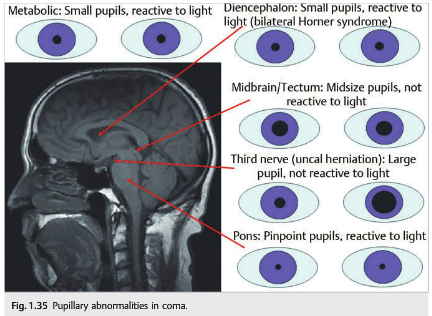
● Pupils (▶Fig. 1.35): Metabolic causes of coma usually cause small reactive pupils. Many toxins and drugs administered may also have effects on the size of the pupils, and pharmacologic mydriasis can inadvertently occur in patients treated with aerosols after extubation.
○ Size
○ Shape
○ Reactivity to light
● Eye position
○ Dysconjugate ocular deviation:– Horizontal, vertical, or oblique misalignment– Often indicates a cranial nerve palsy or skew deviation
○ Conjugate ocular deviation; conjugate lateral eye deviation indicates the following:
– Large lesion in the ipsilateral frontal lobe (the patient looks at his or her lesion)
– Lesion in contralateral pons (gaze palsy) (the patient looks away from the lesion)
– Seizure focus in the contralateral cerebral hemisphere
– Thalamic lesion, which may cause “wrong-way eyes,” with horizontal eye deviation paradoxically looking away from the lesion
● Spontaneous eye movements
○ Roving eye movements: slow ocular conjugate deviations in random directions
– Indicate intact ocular motility function in the brainstem
○ Periodic alternating (“ping-pong”) gaze: slow, repetitive, rhythmic, back-and-forth, horizontal conjugate eye movements
– Indicates intact ocular motility function in the brainstem
○ Spontaneous nystagmus (unusual in coma)
○ Ocular bobbing: conjugate eye movement beginning with a fast downward movement followed by a slow drift back to the midline (similar to a fish bob in the water)– Severe pontine lesion
○ Ocular dipping: slow downward movement followed by a quick upward deviation– Severe pontine lesion
If there are no spontaneous eye movements, perform oculocephalic maneuvering (eliciting eye movements by turning the head horizontally, then vertically). The eyes should deviate in the direction opposite the head turn. This maneuver should not be performed in trauma patients with possible cervical spine injury. If there are no oculocephalic eye movements, test the VOR with the caloric test (apply ice-cold water against the tympanic membranes). The cold water creates convection currents in the endolymph of the horizontal semicircular canals and inhibits the ipsilateral vestibular system. A normal caloric response in an awake patient is when the eyes move slowly toward the irrigated ear, followed by a fast corrective phase to reset the eyes. With warm water, the eyes move slowly away from the irrigated ear (fast phase is toward the irrigated ear). A way to remember this is with the mnemonic COWS: cold–opposite; warm–same. Bilateral caloric stimulation with cold water produces a downward slow phase. Bilateral caloric stimulation with warm water produces an upward slow phase.
Caloric stimulation involves the following steps:
1. Angle the patient’s head at 30 degrees to align the horizontal semicircular canal perpendicular to the floor.
2. Inspect the tympanic membrane (otoscopy) to exclude rupture or cerumen impaction.
3. Irrigate 30 to 60 mL of ice water into the external auditory canal using a large syringe and tubing from a butterfly catheter (without the needle); place a basin under the ear to collect the water.
4. The other ear can be tested after a few minutes.
In the comatose patient, cold caloric stimulation indicates a normal brainstem or bilateral hemispheric dysfunction when there is an ipsilateral tonic slow phase. It indicates a complete brainstem injury when there are no slow or fast eye movements.
Corneal reflex may be absent if there is pontine dysfunction.
Funduscopic examination of the comatose patient is usually performed undilated because pupil monitoring may be important in coma. Bilateral disc edema suggests raised intracranial pressure and should raise the possibility of an intracranial mass or hemorrhage, hydrocephalus, cerebral venous thrombosis, or meningitis. Vitreous hemorrhage (uni- or bilateral) suggests Terson syndrome (related to acutely raised intracranial pressure, most commonly with subarachnoid hemorrhage).
1.17 Summary of the Neuro-ophthalmic Examination ▶Table 1.6 provides a summary of the neuro-ophthalmic examination and can be used as a checklist at bedside.


Reference: 1. Neuro-ophthalmology Illustrated-2nd Edition. Biousse V and Newman NJ. 2012. Thieme
These questions are archived at https://neuro-ophthalmology.
Follow https://twitter.com/
Please send feedback, questions, and corrections to tcooper@stanford.edu.