Questions:
25. Multiple sclerosis is more common in:
a. Men or Women,
b. African-Americans, Caucasians, or Hispanics?
26. What is Lhermitte sign and is it a classic finding in Multiple sclerosis?
27. What are common eye manifestations of multiple sclerosis?
28. What is the 15-year risk of multiple sclerosis after an initial episode of optic neuritis:
a. Overall,
b. With no MRI lesions,
c. With 1 or more lesions on MRI?
29. How common is mild-to-severe eye pain in optic neuritis?
30. What are the characteristics of Neuromyelitis Optica (Devic disease)?
31. What treatment is useful for Neuromyelitis Optica?
________________________________________
Questions with answers?
25. Multiple sclerosis is more common in:
a. Men or Women,
b. African-Americans, Caucasians, or Hispanics?
Multiple sclerosis is more common in women and Caucasians.
26. What is Lhermitte sign and is it a classic finding in Multiple sclerosis?
Lhermitte sign is an electric shock-like sensation that runs down the back and into the limbs when the neck is flexed. The sign suggests a lesion of the dorsal columns of the cervical cord or of the caudal medulla. Although considered a classic finding in multiple sclerosis, it can be caused by a number of conditions.
27. What are common eye manifestations of multiple sclerosis?
Optic neuritis, unilateral or bilateral internuclear ophthalmoplegia, wall-eyed bilateral INO, and acquired pendular nystagmus.
28. What is the 15-year risk of multiple sclerosis after an initial episode of optic neuritis:
a. Overall 50% developed MS
b. With no MRI lesions, 25% developed MS
c. With 1 or more MRI lesions, 72% developed MS?
29. How common is mild-to-severe eye pain in optic neuritis?
In the ONTT, mild-to-severe eye pain present in 92% of patients.
30. What are the characteristics of Neuromyelitis Optica (Devic disease)?
1. Severe, immune-mediated demyelination and axonal damage, predominantly targeting optic nerves and the spinal cord
2. Associated with a serum NMO-IgG antibody that selectively binds aquaporin-4 (AQP4),
3. Present as bilateral or rapidly sequential optic neuritis (leading to visual loss) or transverse myelitis (often causing limb weakness and bladder dysfunction) or both with a typically relapsing course.
31. What treatment is useful for Neuromyelitis Optica?
Treat acute attacks as soon as possible with high-dose intravenous methylprednisolone: 1 gram daily for three to five consecutive days. Often an immunosuppressive agent, such as azathioprine, methotrexate, or mycophenolate is used. In some cases, rituximab, soon after the acute attack and usually be treated for about 5 years after the attack. Consider plasma exchange. No interferon beta drugs! Do worse if used.
The information below is from: Neuro-ophthalmology Illustrated-2nd Edition. Biousse V and Newman NJ. 2012. Theme
20.4 Multiple Sclerosis
Multiple sclerosis (MS) is a demyelinating disease affecting the white matter of the central nervous system (brain, optic nerves, and spinal cord). Young women are more commonly affected. The disease is more common in Caucasians than in other races.
20.4.1 Patient Evaluation
MS present with relapsing – remitting neurological and visual symptoms and signs.
Common Symptoms and Signs of Multiple Sclerosis
The most common symptoms and signs of MS include the following:
● Optic neuritis (often with Uhthoff Phenomenon)
● Vertigo
● Trigeminal neuralgia
● Diplopia
● Sensory disturbances
● Cerebellar syndrome with ataxia
● Lhermitte sign (electric shock-like sensation down the back when flexing the neck)
● Spasticity and weakness
● Urinary incontinence
Symptoms are often multiple, and they come and go over days to weeks. There are no systemic manifestations of MS (no other organ than the central nervous system is affected). Headaches are uncommon as are seizures (only the white matter is typically affected). Altered mental status is uncommon.
Pearls
The association of neurologic signs with other organ failure, headaches, altered mental status, or seizures, should suggest a diagnosis other than MS, such as vasculitis.
Neuro-ophthalmic Manifestations of Multiple Sclerosis
Visual symptoms and signs are very common and include the following:
● Optic neuritis is very common
○ Posterior (retrobulbar) in 65%
○ Anterior (with disc edema) in 35%
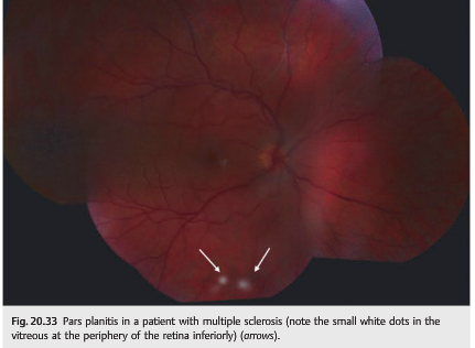
● Uveitis (most often pars planitis) is rare
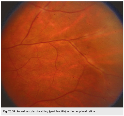
● Retinal periphlebitis (asymptomatic)
● Visual field defects (optic tract lesion)
● Cranial nerve palsies (sixth, third, or fourth nerve palsy)
● Vertigo is very common
● Nystagmus is very common
● Internuclear ophthalmoplegia (bilateral>unilateral) is very common
● Skew deviation
Pearls
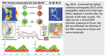
Optic neuritis is often the first sign of multiple sclerosis. Almost all patients with multiple sclerosis develop optic nerve involvement during the course of the disease (▶Fig. 20.31). Optical coherence tomography (OCT) is routinely used to follow patients with MS and is particularly useful in clinical trials (▶Fig. 20.31 e).
Peripheral venous sheathing and pars planitis are usually asymptomatic and found only when a careful dilated funduscopic examination is performed (▶Fig. 20.32 and ▶Fig. 20.33). They are not specific to MS, and they are often found in isolation or in patients with sarcoidosis.
20.4.2 Diagnosis
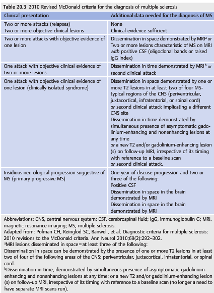
The diagnosis of MS is made when there is evidence of dissemination of the lesions in space (more than one type of deficit or more than one radiologic lesion) and time (relapse of events).
The diagnosis is suspected clinically and is confirmed by brain MRI. When patients present initially with a clinically isolated syndrome (such as optic neuritis), the clinical diagnosis of MS cannot be confirmed until a second clinical event occurs (clinically definite MS). ▶Table 20.3 presents the 2010 revised McDonald criteria for the diagnosis of MS.
20.4.3 Course of the Disease
A relapsing and remitting course is typical: the neurologic symptoms and signs of MS tend to improve spontaneously within a few weeks. They may resolve completely or improve only partially. Each relapse may be associated with a different type of deficit and may leave a residual deficit (neurologic deficit or visual loss) (relapsing-remitting form of MS).In the later course of the disease, the deficits (particularly weakness and spasticity)tend to progress slowly, without remission (secondary progressive form of MS). In a minority of cases, the deficit is slowly progressive from the beginning (primary progressive form of MS).
20.4.4 Treatment of Multiple Sclerosis-Related Neuro-ophthalmic Symptoms
Most MS patients develop visual symptoms at some point during the course of the disease. Monocular or binocular visual loss secondary to recurrent episodes of optic neuritis is common, and numerous patients experience binocular diplopia, often with oscillopsia related to nystagmus. The treatment of these neuro-ophthalmic manifestations of MS is similar to the one recommended for any neurologic flare of MS and is usually administered by the treating neurologist. Symptomatic treatment of persistent visual loss and diplopia is essential and requires a close collaboration with ophthalmologists (see Chapter 21). The treatment of pendular nystagmus is often challenging. It is important for MS patients to be followed by an ophthalmologist who will be able to confirm a diagnosis of optic neuritis when visual loss occurs (and will rule out other causes of visual loss).

The information below is from: Up-to-Date
TREATMENT — Acute attacks and relapses of NMO are generally treated with intravenous glucocorticoids followed soon by therapeutic plasma exchange for refractory or progressive symptoms [116,117]. For prevention of recurrent attacks, treatment with systemic immunosuppression is the mainstay. However, there are no controlled trials evaluating the treatment of NMO, and recommendations are primarily supported by data from observational studies and by the clinical experience of experts.
The rationale for treatment of acute and recurrent attacks in NMO is based upon evidence that humoral autoimmunity plays a role in the pathogenesis of NMO, and is driven by the high attack-related disability, poor prognosis, and overall high risk of mortality in untreated patients [16,118].
Initial and subsequent acute attacks — All patients with suspected NMO should be treated for acute attacks. We suggest initial treatment with high-dose intravenous methylprednisolone (1 gram daily for three to five consecutive days), in agreement with expert panel recommendations and based upon studies of multiple sclerosis and idiopathic optic neuritis [16,119,120]. (See “Treatment of acute exacerbations of multiple sclerosis in adults”, section on ‘Glucocorticoids’ and “Optic neuritis: Prognosis and treatment”, section on ‘Treatment’.).
For patients with severe symptoms, unresponsive to glucocorticoids, therapeutic plasma exchange is the suggested rescue treatment [16,119,120]. Exchanges are carried out every other day up to a total of seven exchanges.
Limited retrospective and uncontrolled data suggest that patients with severe attacks of NMOSD do better if plasma exchange is started early as adjunctive therapy with glucocorticoids. One report of patients with severe attacks (defined by an Expanded Disability Status Scale (table 4) score ≥4 and/or visual acuity ≤20/200) found that initial treatment with intravenous glucocorticoids plus early therapeutic plasma exchange was associated with improved outcomes compared with delayed initiation of plasma exchange following glucocorticoid treatment [121].
Seronegative NMO is managed in the same way as seropositive NMO.
Intravenous immune globulin has not been specifically evaluated for acute attacks of NMO and is rarely used in this setting.
Attack prevention — We recommend initiation of long-term immunosuppression treatment for the prevention of attacks as soon as the diagnosis of NMO is made [16,120,122]. Data on the efficacy of preventive therapies is based upon observational studies. The cornerstone of treatment is systemic immunosuppression with agents including azathioprine [123,124], mycophenolate mofetil [125,126], rituximab [127], methotrexate [128,129], mitoxantrone [130], and oral glucocorticoids [131].
The optimal drug regimen and treatment duration are yet to be determined. Although there is no strict consensus, agents most often considered as first-line monotherapy treatments for NMO are azathioprine, rituximab, and mycophenolate mofetil [59,119,122,132]. Comparative data are scant, but one retrospective, nonrandomized study from two tertiary centers in the United States analyzed relapses among patients with NMO or NMOSD who were treated with azathioprine and concomitant prednisone (n = 32) for at least six months, or mycophenolate (n = 28) for at least six months, or with rituximab (n = 30) for at least one month, and followed up after treatment for at least six months [133]. Treatment with all three agents was associated with significant reductions in annualized relapse rates ranging from 72 to 88 percent compared with baseline. As an example, the annualized relapse rate decreased from 2.26 before azathioprine treatment to 0.63 after treatment, a reduction of 72 percent. Treatment failure was defined as the development of any new central nervous system inflammatory event that occurred despite immunosuppressive treatment; treatment failure rates with these drugs varied from 33 to 53 percent.
Immunosuppression is usually continued for at least five years for patients who are seropositive for aquaporin-4 (AQP4) antibodies, including those presenting with a singleattack, because they are at high risk for relapse or conversion to NMO [116]. However, there is no consensus with regard to the duration of immunosuppressive treatment. Some experts suggest that life-long therapy is appropriate, given the often devastating nature of the disease. Others suggest that the length of immunosuppression should be tailored to the severity of attacks and disability.
Treatment with tocilizumab (an IL-6 receptor antagonist) has been associated with clinical stabilization or improvement in a small number of patients with refractory NMO who failed one or more of the “standard” treatments discussed above [134-138]. Similarly, eculizumab (a complement-inhibiting antibody) treatment of NMO was associated with a significant reduction in attack frequency in a small uncontrolled open-label study, though complicated by meningococcal sepsis with full recovery in 1 of the 14 treated patients [139]. In an observational study of five Chinese patients with highly active NMOSD that was poorly responsive to “standard” immunosuppressive agents, treatment with bortezomib (a proteosome inhibitor that depletes plasma cells) was associated with relapse-free status in four and clinical stabilization in all five [140].
Limited observational evidence suggests that treatment of NMO with interferon beta, natalizumab, or fingolimod is not effective and may be harmful [141-146]. There is no published evidence regarding the treatment of NMO with ocrelizumab.
References:
1. Neuro-ophthalmology Illustrated-2nd Edition. Biousse V and Newman NJ. 2012. Theme
2. Multiple sclerosis risk after optic neuritis: final optic neuritis treatment trial follow-up.
The Optic Neuritis Study Group. Arch Neurol. 2008 Jun; 65(6): 727–732.
Full Text https://www.ncbi.nlm.nih.gov/pmc/articles/PMC2440583/
3. Up-to-Date Neuromyelitis optica spectrum disorders
Authors: Christopher C Glisson, DO, MS, FAAN
Section Editor: Francisco González-Scarano, MD
Deputy Editor: John F Dashe, MD, PhD
Literature review current through: Sep 2018. | This topic last updated: Oct 02, 2018.
These questions are archived at https://neuro-ophthalmology.stanford.edu
Follow https://twitter.com/NeuroOphthQandA to be notified of new neuro-ophthalmology questions of the week.
Please send feedback, questions, and corrections to tcooper@stanford.edu.