Question:
Which of the following are indicative of pseudoedema of the optic disc rather than disc edema?
1. Sharp disc margins
2. Obscured vessels
3. Venous dilation and tortuosity
4. Peripapillary hemorrhages and exudates
5. Leakage on fluorescein angiogram
6. Anomalous retinal vasculature (arterial branching)
 1
1
____________________________________________________
Answers:
1. Sharp disc margins
6. Anomalous retinal vasculature (arterial branching)
Explanation: “9 Disc Edema
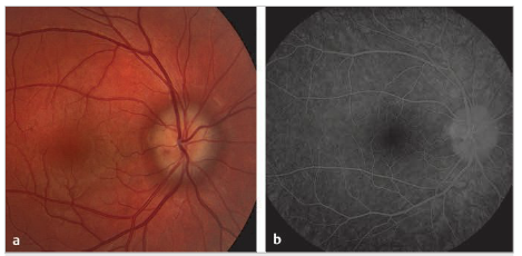
Edema of the optic nerve head, or disc edema, is a nonspecific term describing localized swelling anterior to the lamina cribrosa (▶Fig. 9.1).
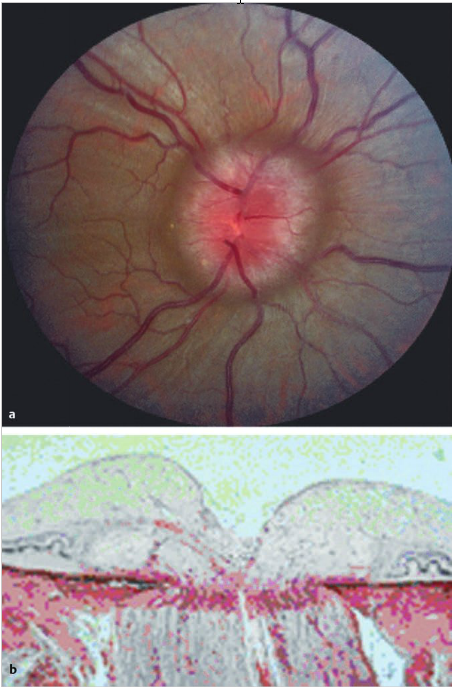
Fig.9.1 (a) Optic nerve swelling in the right eye. The disc margins are blurry, and there is no central cup. (b) Sagittal section of a swollen optic nerve.
9.1 Mechanisms of Optic Nerve Edema
Mechanisms of optic nerve edema include the following:
1. Local optic nerve injury, such as from inflammation (anterior optic neuritis or papillitis), ischemia (anterior ischemic optic neuropathy), fluctuations in intraocular pressure (high, as in acute glaucoma, or low, as in ocular hypotony), and toxicity
2. Blockage of retrograde axonal transport from optic nerve compression (optic nerve tumor or orbital mass) and raised intracranial pressure (papilledema)
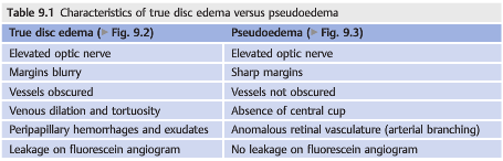
9.2 Differentiating True Disc Edema from Pseudoedema
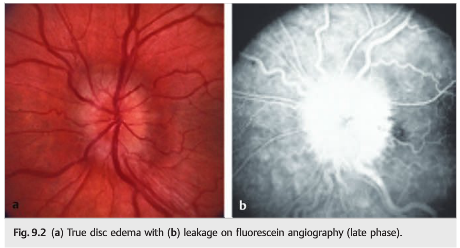
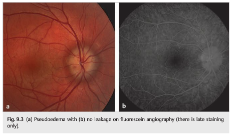
Differentiating true optic nerve head edema from pseudoedema is essential (▶Table 9.1, ▶Fig. 9.2 and ▶Fig. 9.3). In most cases, pseudoedema appearance results from a congenital anomaly of the optic nerve and does not require any workup, whereas true disc edema is associated with numerous concerning disorders.
9.3 Differential Diagnosis of Disc Edema
Disc elevation without true swelling:
● Optic disc anomalies
○ Myelinated nerve fibers
○ Drusen
○ Tilted disc
○ Crowded disc
● Optic disc infiltration
● Leber hereditary optic neuropathy
True disc swelling:
● Elevated intracranial pressure (papilledema)
● Inflammatory optic neuropathy
○ Demyelinating
○ Sarcoidosis or other inflammatory diseases
○ Infectious
● Neuroretinitis
● Vascular optic neuropathy
○ Anterior ischemic optic neuropathy
– Nonarteritic
– Arteritic
○ Diabetic papillopathy
○ Central retinal vein occlusion
○ Carotid-cavernous fistula
○ Malignant systemic hypertension
● Compressive optic neuropathy
○ Neoplastic
– Meningioma
– Hemangioma
– Lymphangioma
○ Non-neoplastic
– Thyroid ophthalmopathy
– Orbital inflammatory pseudotumor
● Infiltrative optic neuropathy
○ Neoplastic
– Leukemia
– Lymphoma
– Glioma
○ Non-neoplastic
– Sarcoidosis
● Toxic
● Metabolic/nutritional deficiencies
● Traumatic optic neuropathy
● Intraocular hypotony (low intraocular pressure)”1
Reference:
1. Neuro-ophthalmology Illustrated-2nd Edition. Biousse V and Newman NJ. 2012. Theme
These questions are archived at https://neuro-ophthalmology.stanford.edu
Follow https://twitter.com/NeuroOphthQandA to be notified of new neuro-ophthalmology questions of the week.
Please send feedback, questions, and corrections totcooper@stanford.edu.