Question:
Which of the following are correct for giant cell arteritis?
1. Visual loss may be preceded by recurrent episodes of transient monocular visual loss.
2. Visual loss may be preceded by recurrent episodes of transient diplopia.
3. AION is its most common ophthalmic manifestation.
4. It is the most common cause of PION.

____________________________________________________
Correct Answers: all are correct.
Explanation:
“8.5.6 Arteritic Anterior and Posterior Ischemic Optic Neuropathy
AION is the most common ophthalmic manifestation of giant cell arteritis, and giant cell arteritis is the most common cause of PION.
Pearls
Suspect and rule out giant cell arteritis in all patients older than age 50 presenting with an ischemic optic neuropathy (ION).
Characteristics
Arteritic ischemic optic neuropathy is most often seen in older Caucasian patients, typically in their 70s and 80s. It is usually associated with systemic symptoms, such as headache, scalp tenderness, jaw claudication, polymyalgia rheumatica, fatigue, and weight loss. However, visual loss can be the only manifestation of the disease (so-called occult forms of giant cell arteritis).
Visual loss in arteritic ION is usually severe, with acuities reduced to no light perception, light perception only, or hand motion. It is often bilateral and can be associated with a concurrent retinal or choroidal infarction. Visual loss may be preceded by recurrent episodes of transient monocular visual loss or transient diplopia.
Diagnosis
Elevation of erythrocyte sedimentation rate (ESR) and C-reactive protein (CRP) is highly suggestive of the disease. The diagnosis is proven by finding granulomatous inflammation with giant cells and disruption of the internal elastic lamina on biopsy of a superficial temporal artery. In the absence of treatment, vision deteriorates, and there is a high risk of involvement of the second eye within days or weeks.
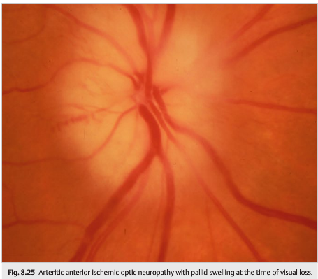
In arteritic AION, the visual loss is profound. The optic nerve appears pale, even acutely, at the time of visual loss. The peripapillary retina is often pallid, suggesting associated choroidal and retinal ischemia. Cotton wool spots are common and indicated use ocular ischemia when associated with AION (▶Fig. 8.25).
Causes
Causes of arteritic AION and PION are giant cell arteritis and systemic vasculitis other than giant cell arteritis, such as systemic lupus erythematosus, periarteritis nodosa, and Churg–Strauss syndrome.
Treatment
Arteritic AION and PION require emergency treatment to prevent complete blindness. Systemic corticosteroid therapy should be instituted immediately upon presumed diagnosis and should not be delayed awaiting results of the temporal artery biopsy.”2
References:
1. Anterior Ischemic Optic Neuropathy. Retina Gallery http://retinagallery.com/displayimage.php?album=281&pid=3199#top_display_media
2. Neuro-ophthalmology Illustrated-2nd Edition. Biousse V and Newman NJ. 2012. Theme
These questions are archived at https://neuro-ophthalmology.stanford.edu
Follow https://twitter.com/NeuroOphthQandA to be notified of new neuro-ophthalmology questions of the week.
Please send feedback, questions, and corrections to tcooper@stanford.edu.