Question:
1. In acute optic neuropathy, how long after onset does the optic nerve head become pale?
2. When is electrophysiologic testing useful in acute optic neuropathies?
3. What does a painful orbital apex syndrome in a diabetic patient suggest?
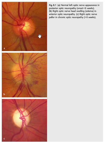
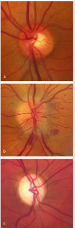 Fig.8.1
____________________________________________________
Correct Answers:
1. In almost all cases of acute optic neuropathy the optic nerve becomes pale 4 to 6 weeks after the onset of visual loss, even when vision recovers.
2. Visual evoked responses and electroretinograms are useful when the diagnosis is unclear (e.g., in cases with bilateral visual loss and no RAPD and when there is concern for a retinal disease rather than optic neuropathy).
3. A painful orbital apex syndrome is highly suggestive of mucormycosis infection.
The information below is from: Neuro-ophthalmology Illustrated-2nd Edition. Biousse V and Newman NJ. 2012. Theme
8 Optic Neuropathies
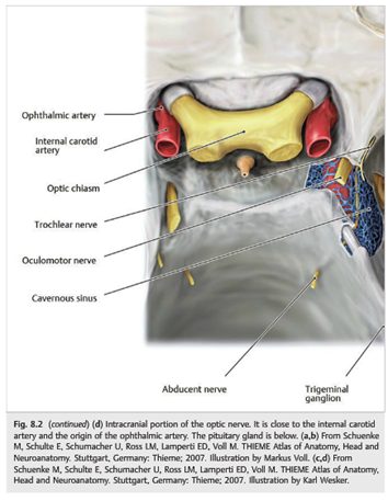
Disorders of the optic nerve are called optic neuropathies. Optic neuropathies associated acutely with a normal optic nerve are referred to as posterior or retrobulbar optic neuropathies. Those with optic nerve head swelling are anterior optic neuropathies. In Almost all cases, the optic nerve becomes pale (optic atrophy) 4 to 6 weeks after the onset of visual loss, even when vision recovers (▶Fig. 8.1 and ▶Fig. 8.2).
8.1 Diagnosis
The diagnosis of optic neuropathy is based on clinical examination, checking for the following:
● Visual loss
● Impaired color vision
● Abnormal visual field
● Relative afferent pupillary defect (RAPD) in all unilateral or asymmetric optic neuropathies (▶Fig. 8.3)
● Optic nerve head appearance
○ Acutely: normal or swollen
○ Late (after 4–6 weeks): pale
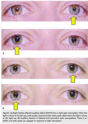
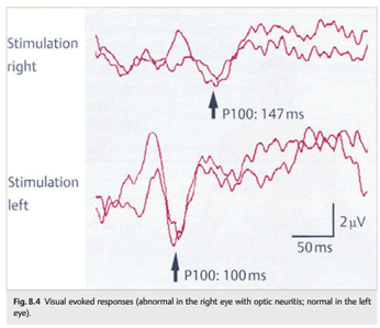
Electrophysiologic testing is most often unnecessary. However, visual evoked responses and electroretinography are useful when the diagnosis is unclear (e.g., in cases with bilateral visual loss and no RAPD and when there is concern for a retinal disease rather than optic neuropathy). Patients with optic neuropathies have abnormal visual evoked responses. The P100 latency is delayed, and the amplitude is decreased (▶Fig. 8.4).
8.1.1 Localization of the Lesion
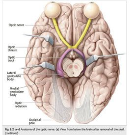
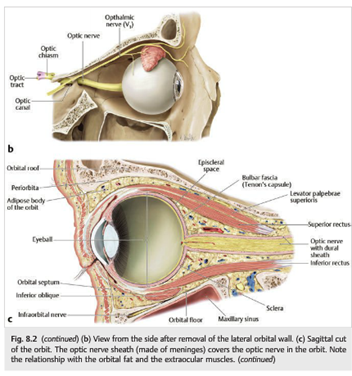
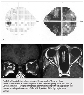
The optic nerve may be affected in the orbit, at the level of the optic canal, or in its intracranial portion. In the orbit, the optic neuropathy may be isolated. The presence of associated symptoms or signs such as diplopia, ptosis, and proptosis suggests a process involving more than just the optic nerve, such as inflammation, infection, or neoplasm (▶Fig. 8.5 and ▶Fig. 8.6)
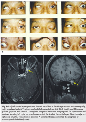
Pearls
A painful orbital apex syndrome in a diabetic patient is highly suggestive of mucormycosis infection.
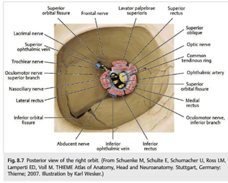
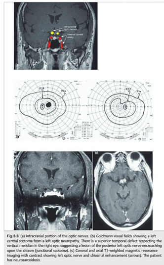
Neurovascular structures enter and exit the orbit through the optic canal and the superior orbital fissure. In the optic canal, the optic nerve exits the orbit, and the ophthalmic artery enters the orbit. In the superior orbital fissure, the superior ophthalmic vein exits the orbit, and cranial nerves III (superior and inferior branches), IV, V1(lacrimal, frontal, and nasociliary nerves), and VI enter the orbit (▶Fig. 8.7). An optic neuropathy may result from a lesion involving the intracranial portion of the optic nerve. When the lesion is close to the optic chiasm, visual field testing demonstrates a junctional scotoma (▶Fig. 8.8).
8.2 Types of Optic Neuropathies (▶Table 8.1)
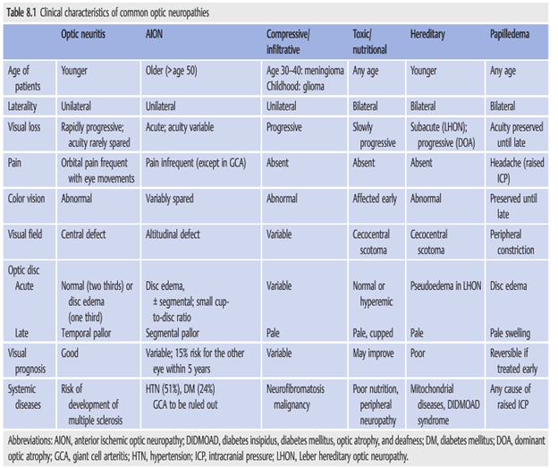
The following lists the main types of optic neuropathies, with subcategories:
- Inflammatory (optic neuritis)
○ Idiopathic demyelinating optic neuritis (associated with multiple sclerosis)
○ Neuromyelitis optica (NMO; Devic disease)
○ Acute disseminated encephalomyelitis
○ Systemic infections
○ Systemic inflammatory diseases (e.g., sarcoidosis) - Vascular (ischemic optic neuropathy)
○ Anterior/posterior
○ Arteritic/nonarteritic - Compressive/infiltrative
○ Neoplastic
○ Non-neoplastic - Hereditary
- Toxic/nutritional
- Traumatic
- Raised intracranial pressure (papilledema)
- Glaucomatous
- Anomalous optic nerve
○ Congenitally anomalous
○ DrusenOptic neuropathies and maculopathies have overlapping presentations. Both cause central visual loss and dyschromatopsia. Many chronic maculopathies are associated with mild optic nerve pallor. When the macula appears normal, it may be difficult to differentiate an optic neuropathy from a maculopathy (▶Table 8.2). Autofluorescence imaging of the macula and spectral optical coherence tomography (OCT) are very helpful in distinguishing optic neuropathies from maculopathies when the macula appears normal on funduscopic examination.
Reference:
1. Neuro-ophthalmology Illustrated-2nd Edition. Biousse V and Newman NJ. 2012. Theme
More than 600 additional neuro-ophthalmology questions are freely available at http://EyeQuiz.com.
Questions prior to September 2016 are archived at http://ophthalmology.stanford.edu/blog/
After that, questions are archived at https://neuro-ophthalmology.stanford.edu
Follow https://twitter.com/NeuroOphthQandA to be notified of new neuro-ophthalmology questions of the week.
Please send feedback, questions and corrections to tcooper@stanford.edu.